Home
Body…
————–
Body is made of many systems:-
- Organ system of human body: It consist of various internal organs and external organs.
- Skeletal system of human body- It consist of Axial Skeleton and Appendicular Skeleton.
- Muscle system of human body-It consist of Smooth muscles, Skeletal muscles and cardiac muscles.
- Endocrine system of human body.
- Nervous system of human body.
- Digestive system of human body.
- Circulatory system
system of human body.
Let’s learn various internal organs of organ system…
HUMAN BODY INTERNAL ORGANS
1. Brain
2. Heart (Detailed)
3. Lungs
4. Kidney
5. Pancreas
6. Liver
7. Stomach
8. Intestines
9. Pituitary gland
10. Blood
Detailed notes on blood
11. Skin
12. Muscle
13. Hair teeth and nails
Let’s learn one by one.
Brain…
————
Brain has two Sides.
Left part of brain control right side and right part of brain control left side.
Parts of brain …
——–
1. Cerebrum—-
Largest part of brain. Made up of cerebral cortex and cerebral medulla called white part and grey part of cerebral muscle. There are various folds and convolution in cerebral muscle.
Function of cerebrum is thinking, making desicions.
2. Cerebellum—-
Starting part of central nervous system .
This is second largest part of brain. It is located behind the cerebrum. Job of cerebellum is control involuntary actions. Function of motor nerves.
It is called little brain. Located in hind part of brain.
Maintain posture and balance.
3. Medulla Oblangatta—-
Helps in control and co-ordination of body.
Functional unit of reflex action.
Looks like a flower , it is located in the base of brain.
It connects brain stem to spinal cord. Cardiovascular and respiration controller.
4. Pineal gland …
Pineal gland is located Just between the eye brows in middle of cerebral knobs. It secrets a juice called Melanie responsible for indusing sleep. It is very small, 2 to 3 inches in size.
5. Pituitary gland…
This is the master of all glands of the body. It is called master gland. It basically doesn’t secrete any juice but gives instructions to thyroid gland and various endocrine glands like liver and pancreas.
Heart…
————-
Heart has four chambers.
Left Artria
Right Ventricle
Left Artria
Left Ventricle
1. Artaries are blood vessels that carry impure blood , de-oxigenated blood.
2. Veins are blood vessels that carry oxygenated blood, pure blood.
3. Pulmonary artery carries de-oxigenated blood to Lungs for Purification.
4. Pulmonary vein carries oxygenated blood to heart.
5. Superior vinacava carries oxygenated blood to upper part of body.
6. Inferior vinacava carries oxygenated blood to lower part of body.
7. Aorta is the Largest blood vessels in the body.
Aorta carries pure blood from left ventricle and supply to all parts of neck and arms.
Aorta also carries oxygenated blood supply throughout the body.
It also carries blood to Kidneys for oxygenated blood is required for filteration.
Aorta is the only artary that carries oxygenated blood.
- Left coronary artery
- Right coronary artery
- Left coronary vein
- Right coronary vein
Are the four blood vessels, of which, artaries carry de-oxigenated blood from hearts left and right chambers to lungs.
Veins carry oxygenated blood to both left and right side of heart from lungs.
72 heartbeats occur in one minute .
Blood capacity of heart is 5 liters.
Cystolic Blood pressure is 80/120 mm .
Lungs…
—————-
Lungs are the organs responsible for breathing.
1.Trachea
Trachea are single tubular structure that are covered by cartialegenus muscle. They send air to lungs from nose.
When air enters from nose, through trachea it enters lungs.
Lungs have three lobes in right side and two lobes in left side. Because pancreas are attached to left side.
2. Bronchus
Trachea branches to Bronchus . Bronchus branch to bronchitis.
3.Alveioli
Bronchitis gets attached to small circular structure called alveioli.
There are artaries and veins dividing into capillary in the border areas of lungs.
These veins have pure oxygenated blood and artaries have de-oxigenated blood.
When air enters alveioli. Blood cells transfer oxygen to them in veins. Artaries supply carbon dioxide into alveioli . We exhale this carbon dioxide through nose .
How does breathing take place?
Lungs are like sponge which remain contracted, this low pressure tends to supply air from outside through nose to lungs. Expanded lungs help blood to extract oxygen to blood cells and expel carbon dioxide outside.
What is respiration?
There are Four types of respiration.
1. BREATHING or ventilation.
2. EXTERNAL RESPIRATION, which is the exchange of gases (oxygen and carbon dioxide) between inhaled air and the blood.
3. INTERNAL RESPIRATION, which is the exchange of gases between the blood and tissue fluids.
4. CELLULAR RESPIRATION.
Cellular respiration is a set of metabolic reactions and processes that take place in the cells of organisms to convert chemical energy from oxygen molecules or nutrients into adenosine triphosphate (ATP), and then release waste products.
Lung capacity is 6 liters in adults.
Kidney…
—————–
Kidney are two bean shaped organs that are connected to urinary bladder.
This kidney function is filtering blood.
1.Nephron…
Nephrons are building-blocks of Kidney.
In stomach, food gets dijested , food is converted to ATP, various unwanted salts and sugar and nitrogenous waste are moved from blood by nephrons.
2. Glomerulas
Nephron have a Bowman’s capsule which is in contact with, glomerulas. Glomerulas is a bunch of blood vessels that carry unwanted salts. Aferrent tubules carry blood into Bowman’s capsule and Eferrent tubules carry filtered blood outside Bowman’s capsule.
Structure of Nephron:- Bowman’s capsule , upper convolution tube, Henrys loop, lower convulsion tube.
3. Bowman’s capsule
Bowman’s capsule is a bowl like structure that stores waste salts and ions from afferent blood vessels.
Salt from glomerulas is separated into Bowman’s capsule and upper convolution tube carries to Henrys loop where again unwanted ions are separated and moved to lower convolution tube leading to Calix and to Urinary tract into urinary bladder and to urethra thereby passing out the urine when smooth muscle in urethra are expanded.
Pancreas…
————————-
Pancreas are seen attached to lower part of Stomach.
Job of pancreas is making digestive juices for easy digestion.
Pancreas is a Endocrine cum Exocrine gland. Meaning, it directly passes hormones into blood as well as into digestive system.
Pancreas release alpha cells, beta cells, delta cells, and f-cells.
Endocrine hormones secreted by Pancreas are
1. Insulin
2. Glycogen
——-
Exocrine hormones secreted by Pancreas are:-
1. ChimeTrypsinogen-
chimotripsin and
2. Trypsinogen-
tripsin are for Protine digestion.
3.Amylase for carbohydrate digestion
4. Lypase for fat digestion
Brush border enzymes-
—-
1.GlucoDexteramylase
2.Maltase
3.Sucrase
4.Lactase
Natural insulin (i.e. insulin released from your pancreas) keeps your blood sugar in a very narrow range. Overnight and between meals, the normal, non-diabetic blood sugar ranges between 60-100mg/dl and 140 mg/dl or less after meals and snacks.
Liver…
————–
Liver has four lobes. Right lobe, left lobe, coudate lobe and inferior lobe. Liver is an exocrine gland.
Means hormons or enzymes secreted by liver are not secreted to blood but Bile is secreted into intestine.
Bile is essentially a lubricant and dijestive fluid. Liver is also an endocrine gland and a blood filter.
Liver is the biggest gland in human body.
Stomach…
————————
Stomach is a bag like structure attached to the food duct esophagus.
The width:lehgth:height is 6:12:6 inches.
Stomach can hold maximum of 2 to 4 liters.
New born can hold only 30ml of milk.
The stomach is a muscular sac located on the left side of the upper abdomen. The stomach receives food from the esophagus. As food reaches the end of the esophagus, it enters the stomach through a muscular valve called the lower esophageal sphincter. The stomach secretes acid and enzymes that digest food.
Intestine…
————————-
There are two intestine. Small intestine and Large intestine.
Small intestine-
………………………..
Small intestine is folded into pallates and grooves. Small intestine is the continuition of stomach. The point of continuition is called intestinal spincher.
Small intestine
5 m long 1 inch width
Parts of small intestine
1.Deodenum
2.Jejunum
3.Illium
Uses various enzymes from liver pancreas and Gallbladder .
Enzymes like Bile from Liver.
Glycotripsin, tripsin, lypase, maltase, amylase from Pancreas.
These enzymes are secreted directly into Deodenum of small intestine.
Large intestine-
………………………..
Large intestine is divided into five.
1.Ceacum
2.Ascending column
3.Transverse column
4.Desending column
5.Rectum
Large intestine is 2 m long and large diameter.
Peristalsis happens contraction of the large intestines for moving forward undigested waste food.
Food is stored in rectum until the waste is expelled outside the body through annus.
Pituitary gland…
Anterior pituitary:-
Somatotrope-somatostanin
Corticotrope-corticotroposin
Thyrotrope-Thyrotropsin
Gonadotrope- Leiutanizing hormone and Follicle stimulating hormone
Lactotrope- Prolatin
Intermediate pituitary:-
Melanin stimulating hormone
Posterior pituitary:-
Antidiuretic hormone or Vassopressin
Oxytocin
Uses:-
Growth
Urin contraction
Child birth
Breast feeding
Sex hormone
Metabolism
Osmoregulation
Thyroid
Temperature
Pain relief
Blood…
A blood cell, also called a hematopoietic cell, hemocyte, or hematocyte, is a cell produced through hematopoiesis and found mainly in the blood. Major types of blood cells include red blood cells (erythrocytes), white blood cells (leukocytes), and platelets (thrombocytes).
Blood cells. Blood contains many types of cells: white blood cells (monocytes, lymphocytes, neutrophils, eosinophils, basophils, and macrophages), red blood cells (erythrocytes), and platelets. Blood circulates through the body in the arteries and veins.
A, B, AB, O each Rh+, and Rh-, total 8 group s
——————————————————————————————-
Leukocytes
What happens if leukocytes are high?
Higher levels of leukocytes in the bloodstream may indicate an infection. This is because WBCs are part of the immune system, and they help fight off disease and infection. Leukocytes may also be found in a urinalysis, or a urine test. High levels of WBCs in your urine also suggest that you have an infection.
What happens if leucocytes are low?
A low WBC count can be serious because it increases your risk of developing a potentially life-threatening infection. Seek prompt medical care if you have a low WBC count and have signs of an infection, such as a fever, swollen lymph nodes, sore throat, or skin lesions.
There are 6 types of Leucocytes.
———————————————————————————————–
What are the Lifespans 6 types of white blood cells and their counts?
Monocytes. They have a longer lifespan than many white blood cells and help to break down bacteria.
Life span:- Normally, circulating monocytes are short-lived and undergo spontaneous apoptosis on a daily basis. Macrophages, however, have a longer life span. The lifespan varies with the “lesion” and “tissue”. In general, tissue resident macrophages are long-lived cells – from more than 3-days to weeks. Again, the life span varies with species. Unlike neutrophils, which are short-lived, macrophages can live for months to years.
High count of Monocytes. A high monocyte count — also called monocytosis — is often associated with chronic or sub-acute infections. It can also be linked with some types of cancer, especially leukemia. A high monocyte count can occur when you are recovering from an acute infection.
Low levels of monocytes tend to develop as a result of medical conditions that lower your overall white blood cell count or treatments for cancer and other serious diseases that suppress the immune system. Causes of low absolute monocyte count include: chemotherapy and radiation therapy, which can injure bone marrow.
————————————————————————————————Lymphocytes. They create antibodies to fight against bacteria, viruses, and other potentially harmful invaders.
High count of Lymphocytes. High lymphocyte blood levels indicate your body is dealing with an infection or other inflammatory condition. Most often, a temporarily high lymphocyte count is a normal effect of your body’s immune system working. Sometimes, lymphocyte levels are elevated because of a serious condition, like leukemia.
LOW COUNT OF LYMPHOCYTES. Lymphocytopenia, also referred to as lymphopenia, occurs when your lymphocyte count in your bloodstream is lower than normal. Severe or chronic low counts can indicate a possible infection or other signficant illness and should be investigated by your doctor. Lymphocytes are a kind of white blood cell.
LIFE SPAN OF LYMPHOCYTES. Types and functions of lymphocytes. Most lymphocytes are short-lived, with an average life span of a week to a few months, but a few live for years, providing a pool of long-lived T and B cells. These cells account for immunologic “memory,” a more rapid, vigorous response to a second encounter with the same antigen.
———————————————————————————————–Neutrophils. They kill and digest bacteria and fungi.
High count of Neutrophils. Having a high percentage of neutrophils in your blood is called neutrophilia. This is a sign that your body has an infection. Neutrophilia can point to a number of underlying conditions and factors, including: infection, most likely bacterial.
LOW COUNT OF NEUTROPHILS. When the body has too few neutrophils, the condition is called neutropenia. This makes it harder for the body to fight off pathogens. As a result, the person is more likely to get sick from infections. In general, an adult who has fewer than 1,000 neutrophils in a microliter of blood has neutropenia.
LIFE SPAN OF NEUTROPHILS. Neutrophils are short-lived cells; their life span from stem cell to removal in the tissues is 12 to 14 days.
———————————————————————————————–Basophils. basophils are responsible for fighting fungal or bacterial infections and viruses. They are a granulocyte cell, which means that they release granules of enzymes to fight against harmful bacteria and germs.
High Count of Basophils. A high count of basophils is called basophilia. This can be caused by hypothyroidism, a condition which occurs when the thyroid gland doesn’t produce enough thyroid hormone. If thyroid hormone is low, it can cause bodily functions to slow down.
LOW COUNT OF BASOPHILS. Often, a low basophils count is related to an allergic reaction which is putting the basophils into overdrive. In these cases, symptoms will include watery eyes, a runny nose, red rash and hives. However, a basophil low can also be caused by a severe allergic anaphylactic reaction.
LIFE SPAN OF BASOPHILS. They are generated from the granulocyte–monocyte progenitors in the bone marrow and populate the periphery as fully mature cells. The lifespan of basophils is short; recently estimated to be in the range of 1–2 days.
———————————————————————————————–Eosinophils. Eosinophils are a type of disease-fighting white blood cell. This condition most often indicates a parasitic infection, an allergic reaction or cancer. You can have high levels of eosinophils in your blood (blood eosinophilia) or in tissues at the site of an infection or inflammation (tissue eosinophilia).
High Count of Eosinophils. A high count could also be caused by an allergic disorder such as asthma, eczema, hay fever, or allergies to substances or certain medications. High eosinophil count can indicate certain autoimmune disorders, Cushing’s disease (a condition caused by heightened cortisol levels), or blood disorders such as leukemia.
LOW COUNT OF EOSIONOPHILS. Low eosinophil counts may also be due to the time of day. Under normal conditions, eosinophil counts are lowest in the morning and highest in the evening. Unless alcohol abuse or Cushing’s disease is suspected, low levels of eosinophils are not usually of concern unless other white cell counts are also abnormally low. As normal levels of eosinophils can be zero, a low level of eosinophils isn’t usually considered a medical problem after one test. However, there are some conditions that can cause a low level of eosinophils, which is known as eosinopenia. An example of this is drunkenness.
LOW SPAN OF EOSIONOPHILS. The tissue life span of eosinophils ranges from 2 to 5 days, depending partly on the tissue studied. However, cytokines increase eosinophil survival in vitro to 14 days or longer; thus, they likely also prolong eosinophil survival in vivo.
————————————————————————————————
Macrophages. Macrophages are specialised cells involved in the detection, phagocytosis and destruction of bacteria and other harmful organisms. In addition, they can also present antigens to T cells and initiate inflammation by releasing molecules (known as cytokines) that activate other cells.
Kupffer cells: Liver
Alveolar macrophage: Lung alveoli
Type of macrophage: Location
Microglia: Central nervous system
What Does a High Monocyte Count Mean?
A high monocyte count — also called monocytosis — is often associated with chronic or sub-acute infections. It can also be linked with some types of cancer, especially leukemia. A high monocyte count can occur when you are recovering from an acute infection.
Low absolute monocyte count.
Low levels of monocytes tend to develop as a result of medical conditions that lower your overall white blood cell count or treatments for cancer and other serious diseases that suppress the immune system.
Life span of Microphages.
The lifespan varies with the “lesion” and “tissue”. In general, tissue resident macrophages are long-lived cells – from more than 3-days to weeks. Again, the life span varies with species. Unlike neutrophils, which are short-lived, macrophages can live for months to years.
Why are macrophages long-lived?
Macrophages are specialised immune cells that destroy bacteria and other harmful organisms. Most importantly, these long-lived macrophages are vital for the survival of the nerve cells of the gastrointestinal tract. This sheds new light on neurodegenerative conditions of the intestine, but also of the brain.
———————————————————————————————–
Thrombocytes or Platelets
Thrombocytes are pieces of very large cells in the bone marrow called megakaryocytes.
They help form blood clots to slow or stop bleeding and to help wounds heal.
Having too many or too few thrombocytes or having platelets that don’t work as they should can cause problems.
LIFE SPAN OF THROMBOCYTES. The blood cells whose function is to prevent bleeding are platelets and they are also called thrombocytes. The average lifespan of the circulating platelets is around 7 to 10 days.
LIFE SPAN OF ERYTHROCYTES.
Erythrocytes and platelets both begin their lives as hematocytoblasts, or stem cells, within the bone marrow.
This is why erythrocytes only have a lifespan of 120 days, while platelets only live for about ten days.
———————————————————————————————-
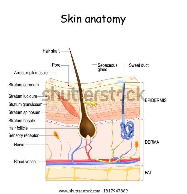
SKIN
The human skin is the outer covering of the body and is the largest organ of the integumentary system. The skin has up to seven layers of ectodermal tissue and guards the underlying muscles, bones, ligaments and internal organs. … Though nearly all human skin is covered with hair follicles, it can appear hairless.
What are the 7 layers of skin?What are the seven most important layers of your skin?
Stratum corneum. The stratum corneum is the outer layer of the skin (epidermis). It serves as the primary barrier between the body and the environment. … stratum corneum: the outermost layer of skin, made up of layers of very resilient and specialized skin cells and keratin.
Stratum lucidum. The stratum lucidum (Latin for “clear layer”) is a thin, clear layer of dead skin cells in the epidermis named for its translucent appearance under a microscope. It is readily visible by light microscopy only in areas of thick skin, which are found on the palms of the hands and the soles of the feet.
Stratum granulosum. The stratum granulosum (or granular layer) is a thin layer of cells in the epidermis lying above the stratum spinosum and below the stratum corneum (stratum lucidum on the soles and palms). Keratinocytes migrating from the underlying stratum spinosum become known as granular cells in this layer.
Stratum spinosum. The stratum spinosum (or spinous layer/prickle cell layer) is a layer of the epidermis found between the stratum granulosum and stratum basale. This layer is composed of polyhedral keratinocytes. These are joined together with desmosomes. … The stratum spinosum also contains Langerhans cells.
Stratum basale. The stratum basale (basal layer, sometimes referred to as stratum germinativum) is the deepest layer of the five layers of the epidermis, the external covering of skin in mammals. The stratum basale is a single layer of columnar or cuboidal basal cells. … The nucleus is large, ovoid and occupies most of the cell.
Dermis. (DER-mis) The inner layer of the two main layers of the skin. The dermis has connective tissue, blood vessels, oil and sweat glands, nerves, hair follicles, and other structures. It is made up of a thin upper layer called the papillary dermis, and a thick lower layer called the reticular dermis.
Hypodermis. The hypodermis is the bottom layer of skin in your body. It has many important functions, including storing energy, connecting the dermis layer of your skin to your muscles and bones, insulating your body and protecting your body from harm. As you age, your hypodermis decreases in size, and your skin starts to sag.
Epidermis. Your epidermis is the top layer of the skin that you can see and touch. Keratin, a protein inside skin cells, makes up the skin cells and, along with other proteins, sticks together to form this layer.
1.What are the 3 major epidermis made up of?
keratinocytes. Three main populations of cells reside in the epidermis: keratinocytes, melanocytes, and Langerhans cells.
The cutaneous membrane is the technical term for our skin. … Our skin is made of three general layers. In order from most superficial to deepest they are the epidermis, dermis, and subcutaneous tissue.
2.What does Skin secrete?
Skin secretions originate from glands that in dermal layer of the epidermis. Sweat, a physiological aid to body temperature regulation, is secreted by eccrine glands. Sebaceous glands secrete the skin lubricant sebum. Sebum is secreted onto the hair shaft and it prevents the hair from splitting.
3.What is the deepest layer of the skin?
hypodermis.
The hypodermis is deep to the dermis and is also called subcutaneous fascia. It is the deepest layer of skin and contains adipose lobules along with some skin appendages like the hair follicles, sensory neurons, and blood vessels.
4.What are the functions of Skin?
The skin performs six primary functions which include, protection, absorption, excretion, secretion, regulation and sensation. The skin functions as our first line of defense against toxins, radiation and harmful pollutants.
Your skin is the organ that comes into contact with the rest of the world. It holds body fluids in, preventing dehydration (dee-hahy-DREY-shun), and keeps harmful microbes (MYE-krobs) out—without it, we would get infections. Your skin is full of nerve endings that help you feel things like heat, cold, and pain.
5.What gives skin its color?
Your skin gets its color from a pigment called melanin. Special cells in the skin make melanin. When these cells become damaged or unhealthy, it affects melanin production. Some pigmentation disorders affect just patches of skin.
——————————————————————————————
MUSCLE
Muscle is contractile tissue grouped into coordinated systems for greater efficiency. In humans the muscle systems are classified by gross appearance and location of cells. The three types of muscles are striated (or skeletal), cardiac, and smooth (or nonstriated).
striated (or skeletal)
Unlike smooth muscle and cardiac muscle, skeletal muscle is under voluntary control. … Similar to cardiac muscle, however, skeletal muscle is striated; its long, thin, multinucleated fibres are crossed with a regular pattern of fine red and white lines, giving the muscle a distinctive appearance.
cardiac muscle
Cardiac muscle (also called heart muscle or myocardium) is one of three types of vertebrate muscle tissue, with the other two being skeletal muscle and smooth muscle. It is involuntary, striated muscle that constitutes the main tissue of the wall of the heart.
smooth (or nonstriated)
Smooth muscle is non-striated, although it contains the same myofilaments they are just organized differently, and involuntary. … Muscle Types: Cardiac and skeletal muscle are both striated in appearance, while smooth muscle is not. Both cardiac and smooth muscle are involuntary while skeletal muscle is voluntary.
What are Myofibrils?
myofibril, very fine contractile fibres, groups of which extend in parallel columns along the length of striated muscle fibres. The myofibrils are made up of thick and thin myofilaments, which help give the muscle its striped appearance.
———————————————————————————————
hair teeth and nails
1. What do hair teeth and nails have in common?
Incisors, hair follicles and nails share a common tissue origin as ectodermal derivatives, and they undergo similar morphogenetic programs. These developmental parallels likely account for some of the similarities in their SC niches.
2. What are 4 types of SC niches?
Hematopoietic stem cell niche.
Hair follicle stem cell niche.
Intestinal stem cell niche.
Cardiovascular stem cell niche.
3.What is morphogenesis?
Morphogenesis is the biological process that causes a cell, tissue or organism to develop … programs according to the spatial patterning of cells within tissues.
In the developmental biology of the early twentieth century, a morphogenetic field is a group of cells able to respond to discrete, localized biochemical
4.What are morphogenetic movements?
The movement of cells in the early embryo that change the shape or form of differentiating cells and tissues.
5.Are hair nails and teeth made of the same thing?
Hair and Fingernails – Like hair and fingernails, tooth enamel contains keratin, but in significantly less levels, teeth are not considered the same makeup as hair or fingernails.
6.Which vitamin is good for hair and teeth?
Vitamin D is doubly important because not only does it boost mineral density, it also helps absorb, carry, and deposit calcium in the bones that support your teeth. Some dairy products and cereal are fortified with vitamin D, but you can also get it naturally from the sun.
The vertebrate ectoderm gives rise to organs that produce mineralized or keratinized substances, including teeth, hair, and claws. Most of these ectodermal derivatives grow continuously throughout the animal’s life and have active pools of adult stem cells that generate all the necessary cell types. These organs provide powerful systems for understanding the mechanisms that enable stem cells to regenerate or renew ectodermally derived tissues, and remarkable progress in our understanding of these systems has been made in recent years using mouse models. We briefly compare what is known about stem cells and their niches in incisors, hair follicles, and claws, and we examine expression of Gli1 as a potential example of a shared stem cell marker. We summarize some of the features, structures, and functions of the stem cell niches in these ectodermal derivatives; definition of the basic elements of the stem cell niches in these organs will provide guiding principles for identification and characterization of the niche in similar systems.
7. Why are hair and nails part of the integumentary system?
The integumentary system is made up of several organs and structures including the skin, hair, nails, glands, and nerves. The primary function of the integumentary system is to protect the inside of the body from elements in the environment—like bacteria, pollution, and UV rays from the sun.
———————————————————————————————
———————————————————————————————–
cell
A cell is the building blocks of all organisms, the smallest unit of a living thing. There are organisms made up of just one cell such as bacteria. And then organisms such as humans that have about 100 trillion cells!
What are the 4 types of cells?
The Four Main Types of Cells
Epithelial Cells. These cells are tightly attached to one another. …
Epithelial cells are a type of cell that lines the surfaces of your body. They are found on your skin, blood vessels, urinary tract, and organs. An epithelial cells in urine test looks at urine under a microscope to see if the number of your epithelial cells is in the normal range.
The most common cause of epithelial cells in urine is improper urine sample collection. Your doctor may, therefore, ask you to take another urine test. The presence of epithelial cells in urine may indicate infections, kidney disease, or (very rarely) a serious illness such as a tumor.
They perform a variety of functions that include protection, secretion, absorption, excretion, filtration, diffusion, and sensory reception. The cells in epithelial tissue are tightly packed together with very little intercellular matrix.
There are three principal shapes of epithelial cell: squamous, columnar, and cuboidal.
Where Are Epithelial Cells Found? Epithelial cells line the major cavities of the body. Epithelia form the structure of the lung, including the alveoli or air sacs where gas exhange occurs. Cells line most organs, such as the stomach and small intestine, kidney, and pancreas.
Is an epithelial cell a skin cell?Even if you think your skin is one smooth surface, it is actually made of millions of epithelial cells that are tightly packed next to each other. That’s not the only place you find these cells. Epithelial cells also line the inside of your throat, intestines, blood vessels, and all your organs.
Nerve Cells. These cells are specialized for communication. …
Each nerve cell consists of the cell body, which includes the nucleus, a major branching fiber (axon) and numerous smaller branching fibers (dendrites). … The myelin sheath is fatty material that covers, insulates and protects nerves of the brain and spinal cord.
Nerve cells (neurones) are ‘excitable’ cells which can transduce a variety of stimuli into electrical signals, continuously sending information about the external and internal environment (in the form of sequences of action potentials) to the central nervous system (CNS).
Neurons are the cells that make up the brain and the nervous system. … For the spinal cord though, we can say that there are three types of neurons: sensory, motor, and interneurons.
Sensory neurons are the nerve cells that are activated by sensory input from the environment – for example, when you touch a hot surface with your fingertips, the sensory neurons will be the ones firing and sending off signals to the rest of the nervous system about the information they have received. A sensory neuron transmits impulses from a receptor, such as those in the eye or ear, to a more central location in the nervous system, such as the spinal cord.
Motor neurons (MNs) are neuronal cells located in the central nervous system (CNS) controlling a variety of downstream targets. This function infers the existence of MN subtypes matching the identity of the targets they innervate. A motor neuron is a neuron whose cell body is located in the motor cortex, brainstem or the spinal cord, and whose axon (fiber) projects to the spinal cord.
Interneurons are the central nodes of neural circuits, enabling communication between sensory or motor neurons and the central nervous system (CNS). They play vital roles in reflexes, neuronal oscillations, and neurogenesis in the adult mammalian brain.
Based on shapes, neurons are classified into five types namely Unipolar neurons, Bipolar neurons, Pseudounipolar neurons, Anaxonic neurons, and Multipolar neurons.
A unipolar neuron is a neuron in which only one process, called a neurite, extends from the cell body. The neurite then branches to form dendritic and axonal processes. Most neurons in the central nervous systems of invertebrates, including insects, are unipolar. … The axon then splits into two branches. A bipolar neuron, or bipolar cell, is a type of neuron that has two extensions (one axon and one dendrite). Many bipolar cells are specialized sensory neurons for the transmission of sense. As such, they are part of the sensory pathways for smell, sight, taste, hearing, touch, balance and proprioception. A pseudounipolar neuron is a type of neuron which has one extension from its cell body. This type of neuron contains an axon that has split into two branches; one branch travels to the peripheral nervous system and the other to the central nervous system. An anaxonic neuron is a type of neuron where there is no axon or it cannot be differentiated from the dendrites. Being loyal to the etymology of anaxonic. A multipolar neuron is a type of neuron that possesses a single axon and many dendrites (and dendritic branches), allowing for the integration of a great deal of information from other neurons. These processes are projections from the neuron cell body. … Peripherally, multipolar neurons are found in autonomic ganglia.
Muscle Cells. These cells are specialized for contraction. …
What is in a muscle cell?The muscle cell is comprised of myofibrils, each consisting of repeated sections of sarcomeres. The cytoplasm of the muscle cell is called sarcoplasm. The smooth endoplasmic reticulum of the muscle cell is called sarcoplasmic reticulum. The plasma membrane of the muscle cell is termed sarcolemma.
Connective Tissue Cells.
The common cell types in connective tissue include: fibroblasts, mast cells, plasma cells, macrophages, adipocytes, and leukocytes.
Fibroblasts are the most common cell type of connective tissue. They produce both fibers and amorphous ground substance.
A fibroblast is a type of biological cell that synthesizes the extracellular matrix and collagen, produces the structural framework (stroma) for animal tissues, and plays a critical role in wound healing. Fibroblasts are the most common cells of connective tissue in animals.
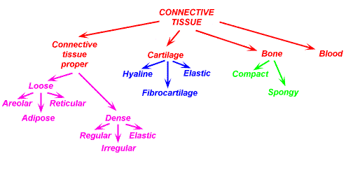
mast cells– A mast cell (also known as a mastocyte or a labrocyte) is a resident cell of connective tissue that contains many granules rich in histamine and heparin. Specifically, it is a type of granulocyte derived from the myeloid stem cell that is a part of the immune and neuroimmune systems.
plasma cells- plasma cell, short-lived antibody-producing cell derived from a type of leukocyte (white blood cell) called a B cell. B cells differentiate into plasma cells that produce antibody molecules closely modeled after the receptors of the precursor B cell.
macrophages- Macrophages are specialised cells involved in the detection, phagocytosis and destruction of bacteria and other harmful organisms. In addition, they can also present antigens to T cells and initiate inflammation by releasing molecules (known as cytokines) that activate other cells.
adipocytes– Adipocytes, also known as lipocytes and fat cells, are the cells that primarily compose adipose tissue, specialized in storing energy as fat. … In cell culture, adipocytes can also form osteoblasts, myocytes and other cell types.
OSTEOBLASTS are the cells that form new bone. They also come from the bone marrow and are related to structural cells. They have only one nucleus. Osteoblasts work in teams to build bone. They produce new bone called “osteoid” which is made of bone collagen and other protein.
MYCOCYTES – Myocytes are contractile muscle cells that make up the heart muscle: • They include one to four nuclei per cell. • They have high mitochondrial organelle density within the cell that can produce ATP for energy.
leukocytes- Leukocytes are part of the body’s immune system. They help the body fight infection and other diseases. Types of leukocytes are granulocytes (neutrophils, eosinophils, and basophils), monocytes, and lymphocytes (T cells and B cells).
NEUTROPHILS- A type of white blood cell that is an important part of the immune system and helps the body fight infection. When microorganisms, such as bacteria or viruses, enter the body, neutrophils are one of the first immune cells to respond.
EOSINOPHILS- Eosinophils are a type of disease-fighting white blood cell. This condition most often indicates a parasitic infection, an allergic reaction or cancer. You can have high levels of eosinophils in your blood (blood eosinophilia) or in tissues at the site of an infection or inflammation (tissue eosinophilia).
BASOPHILS- Basophils are a type of white blood cell. Like most types of white blood cells, basophils are responsible for fighting fungal or bacterial infections and viruses. They are a granulocyte cell, which means that they release granules of enzymes to fight against harmful bacteria and germs.
MONOCYTE- A monocyte is a type of white blood cell and a type of phagocyte. Enlarge. Blood cells. Blood contains many types of cells: white blood cells (monocytes, lymphocytes, neutrophils, eosinophils, basophils, and macrophages), red blood cells (erythrocytes), and platelets.
LYMPHOCYTES- Lymphocytes are white blood cells and one of the body’s main types of immune cells. They are made in the bone marrow and found in the blood and lymph tissue. The immune system is a complex network of cells known as immune cells that include lymphocytes.
MACROPHAGES- Macrophages are specialised cells involved in the detection, phagocytosis and destruction of bacteria and other harmful organisms. In addition, they can also present antigens to T cells and initiate inflammation by releasing molecules (known as cytokines) that activate other cells.
ERYTHROCYTES- Red blood cells, also known as erythrocytes, deliver oxygen to the tissues in your body. Oxygen turns into energy and your tissues release carbon dioxide. Your red blood cells also transport carbon dioxide to your lungs for you to exhale.
PLATELETS- Platelets are tiny blood cells that help your body form clots to stop bleeding. If one of your blood vessels gets damaged, it sends out signals to the platelets. The platelets then rush to the site of damage and form a plug (clot) to fix the damage.
What are the 3 functions of platelets?While the primary function of the platelet is thought to be hemostasis, thrombosis, and wound healing through a complex activation process leading to integrin activation and formation of a “core” and “shell” at the site of injury, other physiological roles for the platelet exist including immunity and communication
A normal platelet count ranges from 150,000 to 450,000 platelets per microliter of blood. Having more than 450,000 platelets is a condition called thrombocytosis; having less than 150,000 is known as thrombocytopenia. You get your platelet number from a routine blood test called a complete blood count (CBC).
Foods rich in folate, vitamin B 12, vitamin C, D, K and iron are known to increase the platelet counts.
- Papaya leaf. …
- Wheatgrass. …
- Pomegranate. …
- Pumpkin. …
- Vitamin C rich foods. …
- Raisins. …
- Brussel sprouts. …
- Beetroot.
——————————————————————————————–
——————————————————————————————–
Tissue…
Tissue is a group of cells that have similar structure and that function together as a unit. A nonliving material, called the intercellular matrix, fills the spaces between the cells. … There are four main tissue types in the body: epithelial, connective, muscle, and nervous. Each is designed for specific functions.
