Skelital System

WHAT IS SKELITAL SYSTEM?

The skeletal system includes all of the bones and joints in the body. Each bone is a complex living organ that is made up of many cells, protein fibers, and minerals. The skeleton acts as a scaffold by providing support and protection for the soft tissues that make up the rest of the body.

The skeletal system works as a support structure for your body. It gives the body its shape, allows movement, makes blood cells, provides protection for organs and stores minerals. The skeletal system is also called the musculoskeletal system.

WHAT IS HUMAN SKELITAL SYSTEM?

The human skeleton is the internal framework of the human body. It is composed of around 270 bones at birth – this total decreases to around 206 bones by adulthood after some bones get fused together. The bone mass in the skeleton reaches maximum density around age 21.

  • Bones – rigid structure.
  • Cartilage – soft, cushions the joint.
  • Ligaments – attach bone to bone.
  • Tendon – attach muscle to bone.

WHAT IS THE FUNCTION OF SKELITAL SYSTEM IN HUMAN BODY?

The skeleton serves six major functions: support, movement, protection, production of blood cells, storage of minerals and endocrine regulation.

Protection

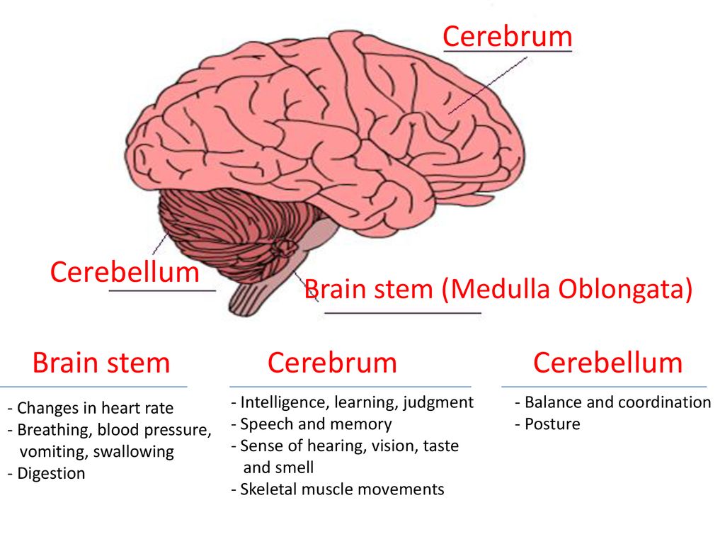
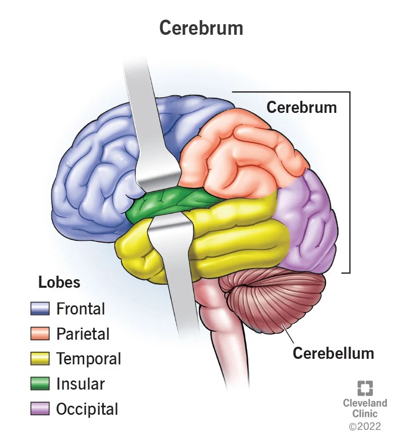
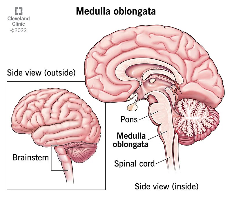
  • The skull protects the brain.
  • The vertebrae protect the spinal cord.
  • The rib cage, spine, and sternum protect the lungs, heart and major blood vessels.

WHICH IS THE BIGGEST BONE IN HUMAN SKELITON?

The femurThe femur is the strongest bone in the body, and it is the longest bone in the human body.

HOW BIG IS THE BIGGEST BONE IN HUMAN SKELITON?

The femur is the longest bone found in the human body. It is almost 19.9 inches long and is commonly known as the thigh bone. One can evaluate that femur is the longest bone just by looking at his/her thigh. It runs from the hip down to around the knee area.

WHICH IS THE SMALLEST BONE IN HUMAN SKELITON?

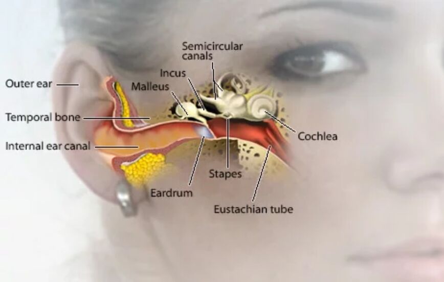
Stapes is ( 3 mm x 2.5 mm ), the “stapes” in the middle ear is the smallest named bone in the human body. The shape of a stirrup, this bone is one of three in the middle ear, collectively known as the ossicles.

WHAT IS THE FUNCTION OF THE SMALLEST BONE IN HUMAN SKELITON?

The stapes bone is essential to our ability to hear. Sounds vibrate the tympanic membrane (the eardrum) and travel through all three bones of the middle ear—the malleus, incus, and stapes.

HUMAN SKELETON

skeleton, Bony framework of the body. It includes the skull, vertebral column, collarbone, shoulder blades, rib cage, pelvic girdle and the bones of the hands, arms, feet, and legs. The skeleton supports the body and protects its internal organs. It is held together by ligaments and moved at the joints by the muscles, which are attached to it. The skeletal system includes both bones and cartilage.

bone, Rigid connective tissue of vertebrates, consisting of cells embedded in a hard matrix. Bones serve as the body’s supporting framework, provide muscle-attachment points for movement, protect the internal organs, house the blood-cell formation system (red bone marrow), and hold about 99% of the calcium vital to many body processes. Bone consists of a matrix of crystals of calcium, chiefly the phosphate and carbonate, embedded among collagen fibres, providing strength and elasticity, and bone cells (less than 5% of its volume). An external layer of compact bone surrounds a central area of spongy bone, except at the marrow cavity. Bone does not grow by cell division; instead, different types of bone cells generate bone matrix, break it down, and maintain it. Bone is remodeled by this process, which strengthens it in areas under greatest stress, permits healing of fractures, and helps regulate calcium levels in body fluid (see calcium deficiency). The process also causes underutilized bone, as in an immobilized limb, to atrophy. Bone disorders include rheumatoid arthritis, osteoarthritis, rickets, osteoporosis, and tumours. Bone can fracture suddenly or over time, as in stress fractures.

cell, in biology, the basic membrane-bound unit that contains the fundamental molecules of life and of which all living things are composed. A single cell is often a complete organism in itself, such as a bacterium or yeast. Other cells acquire specialized functions as they mature. These cells cooperate with other specialized cells and become the building blocks of large multicellular organisms, such as humans and other animals. Although cells are much larger than atoms, they are still very small. The smallest known cells are a group of tiny bacteria called mycoplasmas; some of these single-celled organisms are spheres as small as 0.2 μm in diameter (1μm = about 0.000039 inch), with a total mass of 10−14 gram—equal to that of 8,000,000,000 hydrogen atoms. Cells of humans typically have a mass 400,000 times larger than the mass of a single mycoplasma bacterium, but even human cells are only about 20 μm across. It would require a sheet of about 10,000 human cells to cover the head of a pin, and each human organism is composed of more than 30,000,000,000,000 cells.

osteocyte, a cell that lies within the substance of fully formed bone. It occupies a small chamber called a lacuna, which is contained in the calcified matrix of bone. Osteocytes derive from osteoblasts, or bone-forming cells, and are essentially osteoblasts surrounded by the products they secreted. Cytoplasmic processes of the osteocyte extend away from the cell toward other osteocytes in small channels called canaliculi. By means of these canaliculi, nutrients and waste products are exchanged to maintain the viability of the osteocyte. Osteocytes are the most abundant type of cell in mature bone tissue. They also are long-lived, surviving as long as the bone they occupy exists.

osteoblast, large cell responsible for the synthesis and mineralization of bone during both initial bone formation and later bone remodeling. Osteoblasts form a closely packed sheet on the surface of the bone, from which cellular processes extend through the developing bone. They arise from the differentiation of osteogenic cells in the periosteum, the tissue that covers the outer surface of the bone, and in the endosteum of the marrow cavity. This cell differentiation requires a regular supply of blood, without which cartilage-forming chondroblasts, rather than osteoblasts, are formed. The osteoblasts produce many cell products, including the enzymes alkaline phosphatase and collagenase, growth factors, hormones such as osteocalcin, and collagen, part of the organic unmineralized component of the bone called osteoid. Eventually the osteoblast is surrounded by the growing bone matrix, and, as the material calcifies, the cell is trapped in a space called a lacuna. Thus entrapped, it becomes an osteocyte, or bone cell. Osteocytes communicate with each other as well as with free bone surfaces via extensive cytoplasmic processes that occupy long, meandering channels (canaliculi) through the bone matrix.

————————————————————————————————

skull, skeletal framework of the head of vertebrates, composed of bones or cartilage, which form a unit that protects the brain and some sense organs. The upper jaw, but not the lower, is part of the skull. The human cranium, the part that contains the brain, is globular and relatively large in comparison with the face. In most other animals the facial portion of the skull, including the upper teeth and the nose, is larger than the cranium. In humans the skull is supported by the highest vertebra, called the atlas, permitting nodding motion. The atlas turns on the next-lower vertebra, the axis, to allow for side-to-side motion.

jaw, either of a pair of bones that form the framework of the mouth of vertebrate animals, usually containing teeth and including a movable lower jaw (mandible) and fixed upper jaw (maxilla). Jaws function by moving in opposition to each other and are used for biting, chewing, and the handling of food. The mandible consists of a horizontal arch, which holds the teeth and contains blood vessels and nerves. Two vertical portions (rami) form movable hinge joints on either side of the head, articulating with the glenoid cavity of the temporal bone of the skull. The rami also provide attachment for muscles important in chewing. The centre front of the arch is thickened and buttressed to form a chin, a development unique to man and some of his recent ancestors; the great apes and other animals lack chins.

vertebral column, or spinal column or spine or backbone, Flexible column extending the length of the torso. In humans, it consists of 32–34 vertebrae, with different shapes and functions in each of five regions: 7 cervical, in the neck (including the atlas and axis, modified for free movement of the skull); 12 thoracic, in the chest; 5 lumbar, in the lower back; 5 sacral (fused into the sacrum, part of the pelvic girdle); and 3 to 5 coccygeal (vestigial tailbones fused into the coccyx). The body of each vertebra is separated from its neighbours by cushioning intervertebral disks of cartilage. Behind the body is a Y-shaped vertebral (neural) arch with structures extending up and down to form joints with the adjacent vertebrae and to the back and sides to provide attachment points for muscles and ligaments. The spine supports the torso and protects the spinal cord.

scapula, also called shoulder blade, either of two large bones of the shoulder girdle in vertebrates. In humans they are triangular and lie on the upper back between the levels of the second and eighth ribs. A scapula’s posterior surface is crossed obliquely by a prominent ridge, the spine, which divides the bone into two concave areas, the supraspinous and infraspinous fossae. The spine and fossae give attachment to muscles that act in rotating the arm. The spine ends in the acromion, a process that articulates with the clavicle, or collarbone, in front and helps form the upper part of the shoulder socket. The lateral apex of the triangle is broadened and presents a shallow cavity, the glenoid cavity, which articulates with the head of the bone of the upper arm, the humerus, to form the shoulder joint. Overhanging the glenoid cavity is a beaklike projection, the coracoid process, which completes the shoulder socket. To the margins of the scapula are attached muscles that aid in moving or fixing the shoulder as demanded by movements of the upper limb.

periosteum, dense fibrous membrane covering the surfaces of bones, consisting of an outer fibrous layer and an inner cellular layer (cambium). The outer layer is composed mostly of collagen and contains nerve fibres that cause pain when the tissue is damaged. It also contains many blood vessels, branches of which penetrate the bone to supply the osteocytes, or bone cells. These perpendicular branches pass into the bone along channels known as Volkmann canals to the vessels in the haversian canals, which run the length of the bone. Fibres from the inner layer also penetrate the underlying bone, serving with the blood vessels to bind the periosteum to the bone as Sharpey fibres.

————————————————————————————————

bone formation, also called ossification, process by which new bone is produced. Ossification begins about the third month of fetal life in humans and is completed by late adolescence. The process takes two general forms, one for compact bone, which makes up roughly 80 percent of the skeleton, and the other for cancellous bone, including parts of the skull, the shoulder blades, and the ends of the long bones.

cancellous bone, also called trabecular bone or spongy bone, light, porous bone enclosing numerous large spaces that give a honeycombed or spongy appearance. The bone matrix, or framework, is organized into a three-dimensional latticework of bony processes, called trabeculae, arranged along lines of stress. The spaces between are often filled with marrow and blood vessels. Cancellous bone makes up about 20 percent of the human skeleton, providing structural support and flexibility without the weight of compact bone. It is found in most areas of bone that are not subject to great mechanical stress. It makes up much of the enlarged ends (epiphyses) of the long bones and is the major component of the ribs, the shoulder blades, the flat bones of the skull, and a variety of short, flat bones elsewhere in the skeleton. Cancellous bone is usually surrounded by a shell of compact bone, which provides greater strength and rigidity. The open structure of cancellous bone enables it to dampen sudden stresses, as in load transmission through the joints. Varying proportions of space to bone are found in different bones according to the need for strength or flexibility. Cancellous bone also has a relatively high level of metabolic activity.Cancellous bone can develop into compact bone through the action of bone-forming cells called osteoblasts. It is in that manner that all long bones develop in the embryo. The osteoblasts deposit new bone matrix in layers around the trabeculae, which thus enlarge at the expense of the spaces between them. Eventually the spaces are eliminated, and immature compact bone is produced.

————————————————————————————————

EMBRYONIC SKELETON

Bone of the first type begins in the embryonic skeleton with a cartilage model, which is gradually replaced by bone. Specialized connective tissue cells called osteoblasts secrete a matrix material called osteoid, a gelatinous substance made up of collagen, a fibrous protein, and mucopolysaccharide, an organic glue. Soon after the osteoid is laid down, inorganic salts are deposited in it to form the hardened material recognized as mineralized bone. The cartilage cells die out and are replaced by osteoblasts clustered in ossification centres. Bone formation proceeds outward from these centres. This replacement of cartilage by bone is known as endochondral ossification. Most short bones have a single ossification centre near the middle of the bone; long bones of the arms and legs typically have three, one at the centre of the bone and one at each end. Ossification of long bones proceeds until only a thin strip of cartilage remains at either end; this cartilage, called the epiphyseal plate, persists until the bone reaches its full adult length and is then replaced with bone.

clavicle, also called collarbone, curved anterior bone of the shoulder (pectoral) girdle in vertebrates; it functions as a strut to support the shoulder. The clavicle is present in mammals with prehensile forelimbs and in bats, and it is absent in sea mammals and those adapted for running. The wishbone, or furcula, of birds is composed of the two fused clavicles; a crescent-shaped clavicle is present under the pectoral fin of some fish. In humans the two clavicles, on either side of the anterior base of the neck, are horizontal, S-curved rods that articulate laterally with the outer end of the shoulder blade (the acromion) to help form the shoulder joint; they articulate medially with the breastbone (sternum). Strong ligaments hold the clavicle in place at either end; the shaft gives attachment to muscles of the shoulder girdle and neck.

ear bone, also called Auditory Ossicle, any of the three tiny bones in the middle ear of all mammals. These are the malleus, or hammer, the incus, or anvil, and the stapes, or stirrup. Together they form a short chain that crosses the middle ear and transmits vibrations caused by sound waves from the eardrum membrane to the liquid of the inner ear. The malleus resembles a club more than a hammer, whereas the incus looks like a premolar tooth with an extensive root system. The stapes does closely resemble a stirrup. The top or head of the malleus and the body of the incus are held together by a tightly fitting joint and are seated in the attic, or upper portion, of the eardrum cavity. The handle of the malleus adheres to the upper half of the drum membrane. Three small ligaments hold the head of the malleus, and a fourth attaches a projection (called the short process) from the incus to a slight depression in the back wall of the cavity. The long process of the incus is bent near the lower end and carries a small knoblike bone that is jointed loosely to the head of the stapes—the third and smallest of the ossicles. The stapes lies in a horizontal position at right angles with the long process of the incus. There are two openings in the wall of the bony labyrinth and the stapes footplate fits perfectly in one of these openings—an oval-shaped window, where it is held in place by yet another ligament called the annular ligament. There are two tiny muscles in the middle ear, which serve to alter the tension on the ear bones and thus the intensity (degree of loudness) of sounds. One, the tensor tympani, is attached to the handle of the malleus (itself attached to the eardrum membrane) and by its contraction tends to draw the malleus inward, thus increasing drum membrane tension. The second, called stapedius, tends to pull the footplate of the stapes out of the oval window. This is accomplished by tipping the stirrup, or stapes, backward.

osteoclast, large multinucleated cell responsible for the dissolution and absorption of bone. Bone is a dynamic tissue that is continuously being broken down and restructured in response to such influences as structural stress and the body’s requirement for calcium. The osteoclasts are the mediators of the continuous destruction of bone. Osteoclasts occupy small depressions on the bone’s surface, called Howship lacunae; the lacunae are thought to be caused by erosion of the bone by the osteoclasts’ enzymes. Osteoclasts are formed by the fusion of many cells derived from circulating monocytes in the blood. These in turn are derived from the bone marrow. Osteoclasts may have as many as 200 nuclei, although most have only 5 to 20. The side of the cell closest to the bone contains many small projections (microvilli) that extend into the bone’s surface, forming a ruffled, or brush, border that is the cell’s active region. Osteoclasts produce a number of enzymes, chief among them acid phosphatase, that dissolve both the organic collagen and the inorganic calcium and phosphorus of the bone. Mineralized bone is first broken into fragments; the osteoclast then engulfs the fragments and digests them within cytoplasmic vacuoles. Calcium and phosphorus liberated by the breakdown of the mineralized bone are released into the bloodstream. Unmineralized bone (osteoid) is protected against osteoclastic resorption.

————————————————————————————————

HANDS

carpal bone, any of several small angular bones that in humans make up the wrist (carpus), and in horses, cows, and other quadrupeds the “knee” of the foreleg. They correspond to the tarsal bones of the rear or lower limb. Their number varies. Primitive vertebrates typically had 12. In modern amphibians, reptiles, and birds, the number is reduced by fusion. In humans there are eight, arranged in two rows. The bones in the row toward the forearm are the scaphoid, lunate, triangular, and pisiform. The row toward the fingers, or distal row, includes the trapezium (greater multangular), trapezoid (lesser multangular), capitate, and hamate. The distal row is firmly attached to the metacarpal bones of the hand. The proximal row articulates with the radius (of the forearm) and the articular disk (a fibrous structure between the carpals and malleolus of the ulna) to form the wrist joint.

elbow, in human anatomy, hinge joint formed by the meeting of the humerus (bone of the upper arm) and the radius and ulna (bones of the forearm). The elbow allows the bending and extension of the forearm, and it also allows the rotational movements of the radius and ulna that enable the palm of the hand to be turned upward or downward. The elbow forms from the expansion of the lower end of the humerus into two thick knobs, or condyles: the humerus’ dome-shaped lateral condyle articulates with a shallow depression on the end of the radius, and the humerus’ spool-shaped trochlea fits into a notch in the ulna. In addition, the edge of the radius’ head fits into a shallow groove on the side of the ulna. The bending and extension of the elbow joint are achieved, respectively, by contractions of the biceps and triceps muscles. These movements chiefly involve only the humerus and ulna; rotation of the forearm involves the smaller radius bone as well.

wrist, also called carpus, complex joint between the five metacarpal bones of the hand and the radius and ulna bones of the forearm. The wrist is composed of eight or nine small, short bones (carpal bones) roughly arranged in two rows. The wrist is also made up of several component joints: the distal radioulnar joint, which acts as a pivot for the forearm bones; the radiocarpal joint, between the radius and the first row of carpal bones, involved in wrist flexion and extension; the midcarpal joint, between two of the rows of carpal bones; and various intercarpal joints, between adjacent carpal bones within the rows. The numerous bones and their complex articulations give the wrist its flexibility and wide range of motion. A disk of fibrous cartilage between the radius and the ulna separates the radioulnar joint from the rest of the wrist, which is contained within a capsule of cartilage, synovial membrane, and ligaments. Radiocarpal ligaments carry the hand along with the forearm in rotational movements, and intercarpal ligaments strengthen the small wristbones.

arm, in zoology, either of the forelimbs or upper limbs of ordinarily bipedal vertebrates, particularly humans and other primates. The term is sometimes restricted to the proximal part, from shoulder to elbow (the distal part is then called the forearm). In brachiating (tree-swinging) primates the arm is unusually long.

hand, grasping organ at the end of the forelimb of certain vertebrates that exhibits great mobility and flexibility in the digits and in the whole organ. It is made up of the wrist joint, the carpal bones, the metacarpal bones, and the phalanges. The digits include a medial thumb (when viewed with the palm down), containing two phalanges, and four fingers, each containing three phalanges. {The major function of the hand in all vertebrates except human beings is locomotion; bipedal locomotion in humans frees the hands for a largely manipulative function. In primates the tips of the fingers are covered by fingernails—a specialization that improves manipulation. The palms and undersides of the fingers are marked by creases and covered by ridges called palm prints and fingerprints, which function to improve tactile sensitivity and grip. The friction ridges are arranged in general patterns that are peculiar to each species but that differ in detail. No two individuals are alike, and in humans the patterns are used for identification. The thumb is usually set at an angle distinct from the other digits; in humans and the great apes it rotates at the carpometacarpal joint, and it is therefore opposable to the other fingers and may be used in combination with them to pick up small objects.}

metacarpal, any of several tubular bones between the wrist (carpal) bones and each of the forelimb digits in land vertebrates, corresponding to the metatarsal bones of the foot. Originally numbering five, metacarpals in many mammals have undergone much change and reduction during evolution. The lower leg of the horse, for example, includes only one strengthened metacarpal; the two splint bones behind and above the hoof are reduced metacarpals, and the remaining two original metacarpals have been lost. In humans the five metacarpals are flat at the back of the hand and bowed on the palmar side; they form a longitudinal arch that accommodates the muscles, tendons, and nerves of the palm. The metacarpals also form a transverse arch that allows the fingertips and thumb to be brought together for manipulation.

carpal bone, any of several small angular bones that in humans make up the wrist (carpus), and in horses, cows, and other quadrupeds the “knee” of the foreleg. They correspond to the tarsal bones of the rear or lower limb. Their number varies. Primitive vertebrates typically had 12. In modern amphibians, reptiles, and birds, the number is reduced by fusion. In humans there are eight, arranged in two rows. The bones in the row toward the forearm are the scaphoid, lunate, triangular, and pisiform. The row toward the fingers, or distal row, includes the trapezium (greater multangular), trapezoid (lesser multangular), capitate, and hamate. The distal row is firmly attached to the metacarpal bones of the hand. The proximal row articulates with the radius (of the forearm) and the articular disk (a fibrous structure between the carpals and malleolus of the ulna) to form the wrist joint.

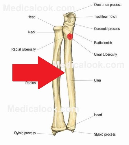
radius, in anatomy, the outer of the two bones of the forearm when viewed with the palm facing forward. All land vertebrates have this bone. In humans it is shorter than the other bone of the forearm, the ulna. The head of the radius is disk-shaped; its upper concave surface articulates with the humerus (upper arm bone) above, and the side surface articulates with the ulna. On the upper part of the shaft is a rough projection, the radial tuberosity, which receives the biceps tendon. A ridge, the interosseous border, extends the length of the shaft and provides attachment for the interosseous membrane connecting the radius and the ulna. The projection on the lower end of the radius, the styloid process, may be felt on the outside of the wrist where it joins the hand. The inside surface of this process presents the U-shaped ulnar notch in which the ulna articulates. Here the radius moves around and crosses the ulna as the hand is turned to cause the palm to face backward (pronation).

ulna, inner of two bones of the forearm when viewed with the palm facing forward. (The other, shorter bone of the forearm is the radius.) The upper end of the ulna presents a large C-shaped notch—the semilunar, or trochlear, notch—which articulates with the trochlea of the humerus (upper arm bone) to form the elbow joint. The projection that forms the upper border of this notch is called the olecranon process; it articulates behind the humerus in the olecranon fossa and may be felt as the point of the elbow. The projection that forms the lower border of the trochlear notch, the coronoid process, enters the coronoid fossa of the humerus when the elbow is flexed. On the outer side is the radial notch, which articulates with the head of the radius. The head of the bone is elsewhere roughened for muscle attachment. The shaft is triangular in cross section; an interosseous ridge extends its length and provides attachment for the interosseous membrane connecting the ulna and the radius. The lower end of the bone presents a small cylindrical head that articulates with the radius at the side and the wrist bones below. Also at the lower end is a styloid process, medially, that articulates with a disk between it and the cuneiform (os triquetrum) wrist bone. The ulna is present in all land vertebrates. In amphibians and some reptiles the radius and ulna do not articulate. The elbow joint evolved first among birds and mammals. The radius tends to be slender in birds; but the ulna is more often reduced in mammals, especially in those adapted for running and, in the case of bats, flying.

The olecranon fossa is a deep triangular depression on the posterior side of the humerus, superior to the trochlea. It provides space for the olecranon of the ulna during extension of the forearm.

The coronoid fossa is a hollow on the anterior surface of the distal end of the humerus, just above the trochlea, in which the coronoid process of the ulna rests when the elbow is flexed.

——————————————————————————————–

LEGS

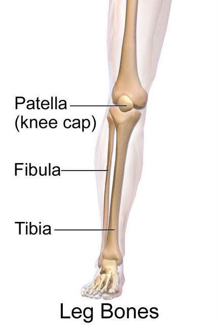
tibia, also called shin, inner and larger of the two bones of the lower leg in vertebrates—the other is the fibula. In humans the tibia forms the lower half of the knee joint above and the inner protuberance of the ankle below. The upper part consists of two fairly flat-topped prominences, or condyles, that articulate with the condyles of the thighbone, or femur, above. The attachment of the ligament of the kneecap, or patella, to the tibial tuberosity in front completes the knee joint. The lateral condyle is larger and includes the point at which the fibula articulates. The tibia’s shaft is approximately triangular in cross section; its markings are influenced by the strength of the attached muscles. It is attached to the fibula throughout its length by an interosseous membrane. At the lower end of the tibia there is a medial extension (the medial malleolus), which forms part of the ankle joint and articulates with the talus (anklebone) below; there is also a fibular notch, which meets the lower end of the shaft of the fibula.

The tibia and fibula are the two long bones in the lower leg. They connect the knee and ankle, but they are separate bones.

ankle, in humans, hinge-type, freely moving synovial joint between the foot and leg. The ankle contains seven tarsal bones that articulate (connect) with each other, with the metatarsal bones of the foot, and with the bones of the lower leg. The articulation of one of the tarsal bones, the ankle bone (talus, or astragalus), with the fibula and tibia of the lower leg forms the actual ankle joint, although the general region is often called the ankle. The chief motions of the ankle are flexion and extension. Like other synovial joints (those joints in which fluid is present), the ankle is subject to such diseases and injuries as bursitis and synovitis.

tarsal, any of several short, angular bones that in humans make up the ankle and that—in animals that walk on their toes (e.g., dogs, cats) or on hoofs—are contained in the hock, lifted off the ground. The tarsals correspond to the carpal bones of the upper limb. In humans the tarsals, in combination with the metatarsal bones, form a longitudinal arch in the foot—a shape well adapted for carrying and transferring weight in bipedal locomotion. In the human ankle there are seven tarsal bones. The talus (astragalus) articulates above with the bones of the lower leg to form the ankle joint. The other six tarsals, tightly bound together by ligaments below the talus, function as a strong weight-bearing platform. The calcaneus, or heel bone, is the largest tarsal and forms the prominence at the back of the foot. The remaining tarsals include the navicular, cuboid, and three cuneiforms. The cuboid and cuneiforms adjoin the metatarsal bones in a firm, nearly immovable joint.

metatarsal, any of several tubular bones between the ankle (tarsal) bones and each of the hindlimb digits, in land vertebrates corresponding to the metacarpal bones of the hand (forepaw). In humans the five metatarsal bones help form longitudinal arches along the inner and outer sides of the foot and a transverse arch at the ball of the foot. The first metatarsal (which adjoins the phalanges of the big toe) is enlarged and strengthened for its weight-bearing function in standing and walking on two feet.

foot, plural feet, in anatomy, terminal part of the leg of a land vertebrate, on which the creature stands. In most two-footed and many four-footed animals, the foot consists of all structures below the ankle joint: heel, arch, digits, and contained bones such as tarsals, metatarsals, and phalanges; in mammals that walk on their toes and in hoofed mammals, it includes the terminal parts of one or more digits.

The major function of the foot in land vertebrates is locomotion. Three types of foot posture exist in mammals: (1) plantigrade, in which the surface of the whole foot touches the ground during locomotion (e.g., human, baboon, and bear), (2) digitigrade, in which only the phalanges (toes and fingers) touch the ground, while the ankle and wrist are elevated (e.g., dog and cat), and (3) unguligrade, in which only a hoof (the tip of one or two digits) touches the ground—a specialization of running animals (e.g., horse and deer).

The human foot is non prehensile and is adapted for a form of bipedalism distinguished by the development of the stride—a long step, during which one leg is behind the vertical axis of the backbone—which allows great distances to be covered with a minimum expenditure of energy. The big toe converges with the others and is held in place by strong ligaments. Its phalanges and metatarsal bones are large and strong. Together, the tarsal and metatarsal bones of the foot form a longitudinal arch, which absorbs shock in walking; a transverse arch, across the metatarsals, also helps distribute weight. The heel bone helps support the longitudinal foot arch.

————————————————————————————————

hip, in anatomy, the joint between the thighbone (femur) and the pelvis; also the area adjacent to this joint. The hip joint is a ball-and-socket joint; the round head of the femur rests in a cavity (the acetabulum) that allows free rotation of the limb. Amphibians and reptiles have relatively weak pelvic girdles, and the femur extends horizontally. This does not permit efficient resistance to gravity, and the trunks of these animals often rest partially on the ground. In mammals the hip joint allows the femur to drop vertically, thus permitting the animal to hold itself off the ground and leading to specializations for running and leaping.

pelvis, also called bony pelvis or pelvic girdle, in human anatomy, basin-shaped complex of bones that connects the trunk and the legs, supports and balances the trunk, and contains and supports the intestines, the urinary bladder, and the internal sex organs.

———————————————————————————————–

Phalanges:

 The bones of the fingers and of the toes. There are generally three phalanges (distal, middle, proximal) for each digit except the thumbs and large toes. The singular of phalanges is phalanx.

Long bones are mostly located in the appendicular skeleton and include bones in the lower limbs (the tibia, fibula, femur, metatarsals, and phalanges) and bones in the upper limbs (the humerus, radius, ulna, metacarpals, and phalanges).

Exocrine Glands

Exocrine glands are glands that secrete substances onto an epithelial surface by way of a duct. Examples of exocrine glands include sweat, salivary, mammary, ceruminous, lacrimal, sebaceous, prostate and mucous.

Sweat Gland

Sweat glands are coiled tubular structures vital for regulating human body temperature. Humans have three different types of sweat glands: eccrine, apocrine, and apoeccrine.

Eccrine sweat glands are abundantly distributed all over the skin and mainly secrete water and electrolytes through the surface of the skin.

Apocrine glands in the skin and eyelid are sweat glands. Most apocrine glands in the skin are in the armpits, the groin, and the area around the nipples of the breast. Apocrine glands in the skin are scent glands, and their secretions usually have an odor.

Apoeccrine glands secrete more sweat than both eccrine and apocrine glands, thus playing a large role in axillary sweating. Apoeccrine glands are sensitive to cholinergic activity, though they can also be activated via adrenergic stimulation. Like eccrine glands, they continuously secrete a thin, watery sweat.

Sweat glands occur all over the body, but are most numerous on the forehead, the armpits, the palms and the soles of the feet. Sweat is mainly water, but it also contains some salts. Its main function is to control body temperature. As the water in the sweat evaporates, the surface of the skin cools.

salivary gland

They produce most of the saliva in your mouth. There are three pairs of major salivary glands: the parotid glands, the submandibular glands, and the sublingual glands.

Most animals have three major pairs of salivary glands that differ in the type of secretion they produce:

  • parotid glands produce a serous, watery secretion.
  • submaxillary (mandibular) glands produce a mixed serous and mucous secretion.
  • sublingual glands secrete a saliva that is predominantly mucous in character.

mammary gland

The mammary gland is a gland located in the breasts of females that is responsible for lactation, or the production of milk. Both males and females have glandular tissue within the breasts; however, in females the glandular tissue begins to develop after puberty in response to estrogen release.

Which hormone is responsible for mammary gland?

The serum concentration of prolactin (PRL) and GH are significantly increased, which activates PRLRs and growth hormone receptors (GHRs) to influence ductal and alveolar development and differentiation. Therefore, GH plays a critical role in mammary gland development and milk production

ceruminous gland

Ceruminous glands are specialized sudoriferous glands (sweat glands) located subcutaneously in the external auditory canal, in the outer 1/3. Ceruminous glands are simple, coiled, tubular glands made up of an inner secretory layer of cells and an outer myoepithelial layer of cells. They are classed as apocrine glands.

Where are ceruminous glands found in the body?

External auditory canal. The human ceruminous glands are located in the cartilaginous two-thirds of the external auditory canal (Perry and Shelley,1955). It has been estimated that there are between 1,000 and 2,000 ceruminous glands in the normal ear.

lacrimal gland

The lacrimal gland is located within the orbit above the lateral end of the eye. It continually releases fluid which cleanses and protects the eye’s surface as it lubricates and moistens it. These lacrimal secretions are commonly known as tears.

The lacrimal gland is a bilobed, tear-shaped gland with the primary function of secreting the aqueous portion of the tear film, thereby maintaining the ocular surface. It is primarily located in the anterior, superotemporal orbit within the lacrimal fossa of the frontal bone.

What happens if lacrimal gland is blocked?

Image result for lacrimal gland

Tear glands and tear ducts

Excess fluid drains through the tear ducts into the nose. When you have a blocked tear duct, your tears can’t drain normally, leaving you with a watery, irritated eye. The condition is caused by a partial or complete obstruction in the tear drainage system.

What is the function of tears?Tears keep your eyes wet and smooth, and help focus light so you can see clearly. They also protect your eyes from infections and irritating things, like dirt and dust. Every time you blink, a thin layer of tears called a “tear film” spreads across the surface of your cornea (the clear outer layer of the eye)

sebaceous gland

A sebaceous gland is a microscopic exocrine gland in the skin that opens into a hair follicle to secrete an oily or waxy matter, called sebum, which lubricates the hair and skin of mammals.

The normal function of sebaceous glands is to produce and secrete sebum, a group of complex oils including triglycerides and fatty acid breakdown products, wax esters, squalene, cholesterol esters and cholesterol. Sebum lubricates the skin to protect against friction and makes it more impervious to moisture.

What can the sebaceous gland cause?

Sebaceous glands secrete an oily substance called sebum. This helps protect the skin from the outside environment. Due to an overgrowth of oil-producing cells, sebum can become trapped inside the gland, causing it to swell and form a bump under the skin. This is known as sebaceous hyperplasia.

Can you pop sebaceous glands?Squeezing sebaceous hyperplasia bumps won’t help, as there’s nothing inside that can be extracted. In fact, doing this can actually cause the bumps to become inflamed or bleed.

Do sebaceous cysts go away?

Sebaceous cysts most often disappear on their own and are not dangerous. As stated, however, they may become inflamed, tender, and even infected. Sometimes sebaceous cysts grow large enough that they may interfere with your everyday life.

Does sebum cause hair loss?The overproduction of sebum causes the hair and skin to become greasy and can lead to various complications such as dandruff and acne. If left untreated, sebum buildup on the scalp can cause more serious symptoms and complications, including hair loss.

prostate gland

The prostate gland is located just below the bladder in men and surrounds the top portion of the tube that drains urine from the bladder (urethra). The prostate’s primary function is to produce the fluid that nourishes and transports sperm (seminal fluid).

The prostate has various functions. The most important is producing seminal fluid, a fluid that is a component of semen. It also plays a role in hormone production and helps regulate urine flow. Prostate problems are common, especially in older men.

mucous gland

Mucous gland, also known as muciparous glands, are found in several different parts of the body, and they typically stain lighter than serous glands during standard histological preparation. Most are multicellular, but goblet cells are single-celled glands.

What is the function of mucous gland?

Mucous glands, which aid in maintaining the water balance and offer protection from bacteria, are extremely numerous in fish skin, especially in cyclostomes and teleosts.

Digestive System

Parts of Dijestive system

  • Mouth
  • Teeth
  • Tongue
  • Pharynx
  • Esophagus
  • Stomach
  • Small intestine
  • Large intestine
  • Rectum
  • Anus

There are four steps in the digestion process: 

Ingestion: Ingestion is the process of taking in food through the mouth. In vertebrates, the teeth, saliva, and tongue play important roles in mastication (preparing the food into bolus). While the food is being mechanically broken down, the enzymes in saliva begin to chemically process the food as well.

The mechanical and chemical breakdown of food: Mechanical digestion involves physically breaking down food substances into smaller particles to more efficiently undergo chemical digestion. The role of chemical digestion is to further degrade the molecular structure of the ingested compounds by digestive enzymes into a form that is absorbable into the bloodstream.

Nutrient absorption: Absorption is a complex process, in which nutrients from digested food are harvested. Absorption can occur through five mechanisms: 

(1) Active transport– Active transport is the process of moving molecules across a cellular membrane through the use of cellular energy. … Active transport is used by cells to accumulate needed molecules such as glucose and amino acids. Active transport powered by adenosine triphosphate (ATP) is known as primary active transport.

Example: uptake of glucose in the intestines in humans and the uptake of mineral ions into root hair cells of plants.

Here are some examples of active transport in animals and humans:

  • Sodium-potassium pump (exchange of sodium and potassium ions across cell walls)
  • Amino acids moving along the human intestinal tract.
  • Calcium ions moving from cardiac muscle cells.
  • Glucose moving in or out of a cell.
  • A macrophage ingesting a bacterial cell.

(2) Passive diffusionPassive diffusion is the process by which molecules diffuse from a region of higher concentration to a region of lower concentration. It is the most important mechanism for passage of drugs through membranes. Lipid soluble drugs penetrate lipid membranes with ease.

Example: For example, oxygen diffuses out of the air sacs in your lungs into your bloodstream because oxygen is more concentrated in your lungs than in your blood.

Simple diffusion is the passive movement of solute from a high concentration to a lower concentration until the concentration of the solute is uniform throughout and reaches equilibrium.

(3)Facilitated diffusion:Facilitated diffusion is the transport of substances across a biological membrane from an area of higher concentration to an area of lower concentration with the help of a transport molecule. Since substances move along the direction of their concentration gradient, chemical energy is not directly required.

Example:A common example of facilitated diffusion is the movement of glucose into the cell, where it is used to make ATP. Although glucose can be more concentrated outside of a cell, it cannot cross the lipid bilayer via simple diffusion because it is both large and polar.

(4) Co-transport (or secondary active transport):In secondary active transport, also known as coupled transport or cotransport, energy is used to transport molecules across a membrane; however, in contrast to primary active transport, there is no direct coupling of ATP.

Example:An example of secondary active transport is the movement of glucose in the proximal convoluted tubule.

(5) Endocytosis:Endocytosis is a cellular process in which substances are brought into the cell. The material to be internalized is surrounded by an area of cell membrane, which then buds off inside the cell to form a vesicle containing the ingested material.

Endocytosis is the process of actively transporting molecules into the cell by engulfing it with its membrane. Endocytosis and exocytosis are used by all cells to transport molecules that cannot pass through the membrane passively. Exocytosis provides the opposite function and pushes molecules out of the cell.

Example: The three main types of exocytosis are phagocytosis, pinocytosis and receptor-mediated endocytosis. Pinocytosis is non-specific. Phagocytosis targets large structures (e.g., bacteria, food particles…) and is not particularly specific. … Exocytotic pathways also deliver membrane proteins made in cells to the cell surface.

Elimination of indigestible food:The food molecules that cannot be digested or absorbed need to be eliminated from the body. The removal of indigestible wastes through the anus, in the form of feces, is defecation or elimination.

Organ system

HOME

Internal Organs

1. Brain
2. Heart
3. Lungs
4. Kidney
5. Pancreas
6. Liver
7. Stomach
8. Intestines
9. Pituitary gland
10. Blood

External Organs

1. Eye
2. Ear
3. Nose
4. Tongue
5. Teeth
6. Bone
7. Skin
8. Hair
9. Nails

Brain

What is the definition of brain?

Brain Anatomy and How the Brain Works | Johns Hopkins Medicine

The brain is a complex organ that controls thought, memory, emotion, touch, motor skills, vision, breathing, temperature, hunger and every process that regulates our body. Together, the brain and spinal cord that extends from it make up the central nervous system, or CNS.

What are the 3 types of the brain?

Brain Basics: Know Your Brain | National Institute of ...

The brain can be divided into three basic units: the forebrain, the midbrain, and the hindbrain. The hindbrain includes the upper part of the spinal cord, the brain stem, and a wrinkled ball of tissue called the cerebellum. The hindbrain controls the body’s vital functions such as respiration and heart rate.

What does our brain do?

Brain and Nervous System (for Teens) - Nemours KidsHealth

The brain controls what you think and feel, how you learn and remember, and the way you move and talk. But it also controls things you’re less aware of — like the beating of your heart and the digestion of your food. Think of the brain as a central computer that controls all the body’s functions.

What are the main parts of human brain?

The brain has three main parts: the cerebrum, cerebellum and brainstem.

What is brain and its parts?

The cerebrum is further divided into four sections or lobes: Frontal lobe: It is associated with parts of speech, planning, reasoning, problem-solving and movements. Parietal lobe: Help in movements, the perception of stimuli and orientation. Occipital lobe: It is related to visual processing.

What is the study of the brain?

Neuroscience, also known as Neural Science, is the study of how the nervous system develops, its structure, and what it does. Neuroscientists focus on the brain and its impact on behavior and cognitive functions.

What is the unit of brain?

The basic unit of the brain is the neuron.

Where is brain located?

skull

The brain is housed inside the bony covering called the cranium. The cranium protects the brain from injury. Together, the cranium and bones that protect the face are called the skull. Between the skull and brain is the meninges, which consist of three layers of tissue that cover and protect the brain and spinal cord.

What is brain made of?

The human brain is primarily composed of neurons, glial cells, neural stem cells, and blood vessels. Types of neuron include interneurons, pyramidal cells including Betz cells, motor neurons (upper and lower motor neurons), and cerebellar Purkinje cells.

What size is my brain?

about 5.5 x 6.5 x 3.6 inches

On average, the human brain measures about 5.5 x 6.5 x 3.6 inches (140 x 167 x 93 mm). It weighs about 3 pounds (1.3 kg).

What is the largest part of the brain?

The cerebrum is the largest component, extending across the top of the head down to ear level. The cerebellum is smaller than the cerebrum and located underneath it, behind the ears toward the back of the head.

What is the biggest part of the brain?

Your cerebrum is the largest part of your brain, and it handles a wide range of responsibilities. Located at the front and top of your skull, it gets its name from the Latin word meaning “brain.” Your cerebrum is instrumental in everything you do in day-to-day life, ranging from thoughts to actions.

What are the 4 major of the brain?

Traditionally, each of the hemispheres has been divided into four lobes: frontal, parietal, temporal and occipital.

What part of the brain is balance?

Cerebellum

Cerebellum. The cerebellum sits at the back of the brain and controls your sense of balance. This allows you to stand up, walk in a straight line, and know if you are standing up or sitting down.

What is a brain type 2?

Brain Type 2 — Spontaneous

The Spontaneous Brain Type tends to be creative, out-of-the-box thinkers who would rather do things on the spur of the moment than have a set schedule. You may struggle with a short attention span, distractibility, disorganization, and impulse control problems.

What are 5 main parts of the brain?

We’re going to talk about these five parts, which are key players on the brain team:

  • cerebrum (say: suh-REE-brum)
  • cerebellum (say: sair-uh-BELL-um)
  • brain stem.
  • pituitary (say: puh-TOO-uh-ter-ee) gland.
  • hypothalamus (say: hy-po-THAL-uh-mus)

What part of the brain is balance?

Cerebellum

Cerebellum. The cerebellum sits at the back of the brain and controls your sense of balance. This allows you to stand up, walk in a straight line, and know if you are standing up or sitting down.

What is the center of the brain called?

Brainstem. This is the middle of the brain. It includes the midbrain, the pons, and the medulla. The brainstem controls movement of the eyes, face, and mouth.

What is disorder of the brain?

Brain disease affects millions worldwide and includes a wide spectrum of diseases and disorders—from stroke and Alzheimer’s to multiple sclerosis, epilepsy, traumatic brain injury, and more.

What is brain scientist?

Brain Science and Cognitive Psychology Explores Our Mental ...

Cognitive psychologists, sometimes called brain scientists, study how the human brain works — how we think, remember and learn. They apply psychological science to understand how we perceive events and make decisions.

How is the human brain designed?

Brains are built over time, from the bottom up.

The basic architecture of the brain is constructed through an ongoing process that begins before birth and continues into adulthood. Simpler neural connections and skills form first, followed by more complex circuits and skills.

Why study the brain?

Why study the brain? - Queensland Brain Institute ...

Unravelling how the brain works will help us understand the basis of human behaviour and actions. While each brain is unique, all healthy human brains share the same basic structures and functions.

Who studies brain diseases?

neurologist

A neurologist is a medical doctor who diagnoses, treats and manages disorders of the brain and nervous system (brain, spinal cord and nerves). A neurologist knows the anatomy, function and conditions that affect your nerves and nervous system. Your nervous system is your body’s command center.

Who is called small brain?

Cerebellum, commonly known as the “little brain” because it resembles a miniature cerebrum, is in charge of balance, movement, and coordination. The cerebellum is a brain region that lies underneath the occipital and temporal lobes of the cerebral cortex.

What protects the brain?

The brain is protected by the bones of the skull and by a covering of three thin membranes called meninges. The brain is also cushioned and protected by cerebrospinal fluid. This watery fluid is produced by special cells in the four hollow spaces in the brain, called ventricles.

What is the smallest of the brain?

Situated just below the thalamus, the hypothalamus is the smallest part of the brain. It plays an important role in the nervous system as well as in the endocrine system. It maintains homeostasis, regulates release of hormones and regulates body temperature.

Which side brain is present?

The brain is divided into symmetrical left and right hemispheres. Each hemisphere is in charge of the opposite side of the body, so your right brain controls your left hand. The right hemisphere also takes in sensory input from your left side and vice versa. The brain is segmented into regions called lobes.

What is brain activity?

Defining Functional Brain Activity

Function can be defined as the ability to perform a given cognitive or physiological task.

Which side of the brain works?

The brain’s left half is primarily responsible for speech and abstract thinking. It also controls the right side of the body. The right side of the brain is responsible for image processing, spatial thinking, and movement in the left side of the body.1

Does brain work 24 hours?

How many hours does the brain work? Brain works 24*7*365 days. The only difference is, it works in different levels- conscious, subconscious & unconscious level. The moment your brain stops working, doctors will declare you brain dead or simply dead.

How powerful is the brain?

The human brain can generate about 23 watts of power (enough to power a lightbulb). All that power calls for some much-needed rest. Adequate sleep helps maintain the pathways in your brain. Additionally, sleep deprivation can increase the build-up of a protein in your brain that is linked to Alzheimer’s disease.

What does the brain eat?

The brain needs energy to work, and its main source of energy is glucose. This simple sugar, found in many foods, is carried in the bloodstream and converted into energy by tissue cells throughout the body.

How many kg is the brain?

about 1.4 kilograms

The human brain weighs approximately about 1.4 kilograms and makes up about 2% of the human body weight. The brain is divided into three regions – Forebrain, midbrain, and hindbrain. The forebrain includes the cerebrum, hypothalamus, and thalamus.

Why are brains different?

Some of this variability in the brain—which correlates with differences in personality, cognitive functions, and susceptibility to various neurological and psychiatric syndromes—is accounted for by genetic differences.

Does blood go to the brain?

The brain receives blood from two sources: the internal carotid arteries, which arise at the point in the neck where the common carotid arteries bifurcate, and the vertebral arteries (Figure 1.20). The internal carotid arteries branch to form two major cerebral arteries, the anterior and middle cerebral arteries.

Does my brain have a limit?

The human mind does have a memory limit. The average adult human brain’s memory capacity is 2.5 million gigabytes. However, it doesn’t run out of storage capacity, per se. A single human brain has many different kinds of memories.

How much oxygen does the brain use?

20%

With the average human brain weighing 1400 g (∼2% of total body weight), it therefore consumes ∼49 ml O2 per minute, or 20% of total body oxygen consumed while at rest (21a, 46, 82) (Table 1). Within the brain, oxygen consumption is highly dynamic and region specific.

www.brainfacts.org (Number of Neurons in Brain of a Human Being)

You have 12 cranial nerve pairs. Each nerve pair splits to serve the two sides of your brain and body. For example, you have one pair of olfactory nerves. One olfactory nerve is on the left side of your brain and one is on the right side of your brain.

What are the 4 types of nerves?

It is conventional, however, to describe nerve types on the basis of their function: motor, sensory, autonomic or cranial.

  • Motor Nerves. …
  • Sensory Nerves. …
  • Autonomic Nerves. …
  • Cranial Nerves.
  • The olfactory nerve
  • The olfactory nerve is the first cranial nerve and is instrumental in our sense of smell. The olfactory nerve contains only afferent sensory nerve fibers and, like all cranial nerves, is paired.
  • The central nervous system (defined as the brain and spinal cord) is usually considered to have seven basic parts: the spinal cord, the medulla, the pons, the cerebellum, the midbrain, the diencephalon, and the cerebral hemispheres 

Which nerve is faster?

Neuroscience For Kids - conduction velocity

A-delta nerve fibers can conduct action potentials as fast as a sprinter in the Olympics. The large A-beta nerve fibers, which transmit information related to touch, can have action potentials traveling near the speeds of airplanes.

faculty.washington.edu (Learn everything about Brain)

What is nerve 7 called?

The facial nerve is the seventh cranial nerve (CN VII). It arises from the brain stem and extends posteriorly to the abducens nerve and anteriorly to the vestibulocochlear nerve.

————————————————————————————————

Respiratory system

Home

Respiratory system
——————————–

Respiratory system function:-
1.Pulmonary ventilation
2.Internal and external respiration
3.Olfactory—smell
4.Phonetation

Upper respiratory system
—————————————
Nasal cavity
Pharynx
Larynx

Lower respiratory system
—————————————
Lung
Rib gage

Trachea
Bronchus
Bronchii
Alveioli

Nasal muscles
————————————

Superior conche
Middle conche
Inferior conche


Nasal Muscle bones
——————————–
Nasal
Frontal
Maxela
Ethmoid
Vomer

Pharynx bones
Nasopharyx
Oropharynx
Larnyngopharynx

Larynx
————
Voice and respiratory tract.
Has bones attached to cartialegenus muscle and help in phoenix.

Trachea

————-

The trachea, also known as the windpipe, is a cartilaginous tube that connects the larynx to the bronchi of the lungs, allowing the passage of air, and so is present in almost all air-breathing animals with lungs. The trachea extends from the larynx and branches into the two primary bronchi.

Process of respiration

———————————–

When we breath in the air, through the opening of nose air enters into nasal cavity.

From Nasal cavity Air passes through
1. Superior conche
2. Middle conche
3. Inferior conche
These are nasal muscles to trachea .

Then through trachea, air passes to lungs.

We know, lungs has Bronchus and branches of it called bronchii . And button like structure called Alveioli.

Alveioli has indirect contact to blood capillaries. This blood capillaries carry oxygen throughout the body. This is called oxygen saturation. With oxymeter we can measure this oxygen saturation level in our body. Oxygen saturation must be above 90 otherwise we have to consult a doctor.

Internal respiration:-

——————————-

In blood , oxygen is supplied to red blood cells and with the help of haemoglobin, synthesis of oxygen takes place. This is called internal respiration or cellular respiration.

External respiration:-

——————————-

The bi-product CO2 is returned to alveioli and through Bronchus it returns to trachea and back to nasal cavity and through nasal opening the air is given out. The process is called external respiration.

Circulatory system

In Circulatory system,

Heart consist of two Artria and two ventricle.

Artary carry oxygenated blood.
Veins carry De-oxigenated blood.

Aiorta is the only big artary that carries oxygenated blood through out the body.

Pulmonary vein carries pure blood from heart to body tissues and then De-oxigenated blood to lungs where as pulmonary artery carries pure blood from lungs to heart.

SVC or Superior vinacava carries oxygenated blood to upper part of body.

IVC or Inferior vinacava carries oxygenated blood to lower part of the body.

Right side of heart has tricuspid valve.
Left side of heart has dicuspid valve.

Heart beats 72 times in one minute.

Heart size is of closed wrist.
Heart weight is 300 grams.

Pacemaker is artificial heart placed after heart transplantation.

The circulatory system

Consists of three independent systems that work together:

the heart (cardiovascular), lungs (pulmonary), and arteries, veins, coronary and portal vessels (systemic).

The system is responsible for the flow of blood, nutrients, oxygen and other gases, and as well as hormones to and from cells.

From the small intestine, the blood gathers food nutrients and delivers them to every cell.

  • Blood. Blood consists of:
  • The heart. The heart pumps blood around the body. …
  • The right side of the heart. …
  • The left side of the heart. …
  • Blood vessels. …
  • Arteries. …
  • Capillaries. …
  • Veins.


Excretary System

Home

Excretary System
————————–
NH3-fish
Urea-amphebians
Uric Acid-birds
NH3+Urea+Uric acid – man

Kidney
———–
115-170 grams
Bean shaped
Outer part-cortex
Inner part-medulla

Nephrons
—————-
Nephrons-structural and functional unit of kidney. Size 3cm

Other escretary system parts
———————————————
Ureter
Urinary bladder
Urethra

Chemical reaction
—————————-
De-Amination cycle -Protien-NH3
Orinthin cycle NH3-Urea
Liver converts NH3 to Urea

Physical reaction
—————————-
Ultra filtration
Re-Absorbtion
Tubular Secretion

Structure of neuron
——————————-
Afferent Artiole-impure blood
Efferent Artiole-pure blood
Glomerulas
Bowman’s capsule- Urea vitamins minerals water.
PCT Proximal Convoluted tubule
Loop of Henley
DCT Distil Convoluted tubule
Collecting duct

Function
————-+
PCT- Carries vitamins.
Loop of Henley- Concentrated blood.
DCT- Blood to Urea.
Collecting duct- Urea+ NH3+Uric acid send to Uriter

Diseases
—————
Uriac- urea mixes with urine
GOUT- urine mixes with
elbow joint fluid

Machine
————-
Dialysis machine:-
Connects to vein of patients.
Sends Dialysis fluid =Water 96% + proteins 3%+ other substances. 1% to veins in body temperature.

Kidney transplantation
———————————–
Can be done ✅ with living or dead person to a healthy person.
48 hours before Brain death.
Most successful

————————————————


Nervous system

The nervous system includes the brain, spinal cord, and a complex network of nerves. This system sends messages back and forth between the brain and the body. The brain is what controls all the body’s functions. The spinal cord runs from the brain down through the back.

Believe it or not, there are over 7 trillion nerves in the human body. All these nerves are part of what’s known as your body’s nervous system. You can think of nerves as your body’s electrical wiring — they transmit signals between your brain, spinal cord, and the rest of your body.

Your nerves are made up of: Axons, cord-like groups of fibers in the center of your nerve. Dendrites, branches that carry electrical impulses. Endoneurium, a layer of connective tissue surrounding axons.

Together with the endocrine system, the nervous system is responsible for regulating and maintaining homeostasis.

  1. Who studies the nervous system?

A neurologist is a medical doctor who diagnoses, treats and manages disorders of the brain and nervous system (brain, spinal cord and nerves). A neurologist knows the anatomy, function and conditions that affect your nerves and nervous system. Your nervous system is your body’s command center.

2. Which is the smallest nerve in human body? Answer:- the trochlear nerve

The smallest nerve in the human body is the trochlear nerve. It allows the superior oblique muscle of the eye to move. It is feasible to look down because of this. The nerve also allows for eye movement toward or away from the nose.

3. What is the largest nerve in the body? Answer:- Sciatic Nerve

The sciatic nerve is the longest, largest nerve in your body. Your sciatic nerve roots start in your lower back and run down the back of each leg. Sciatica is the pain or discomfort if your sciatic nerve gets compressed or pinched.

4. What are nerve cells called? Answer:-Neurons

Neurons, the nerve cells that send and receive signals.

5. What are the four major functions of the nervous system?

Answer:- Reception of general sensory information (touch, pressure, temperature, pain, vibration) 2. Receiving and perceiving special sensations (taste, smell, vision, sounds) 3.Integration of sensory information from different parts of the body and processing them. 4. Response generation.

ANATOMY OF NERVOUS SYSTEM:-Neuroanatomy

NEUROSCIENCE:-

Neuroscience describes the scientific study of the mechanics of the central nervous system such as its structure, function, genetics and physiology as well as how this can be applied to understand diseases of the nervous system.

NEUROLOGY:-

Neurology is a specialized area of medicine that concerns disorders and diseases of the nervous system ranging from Alzheimer’s disease through to infection and personality disorders. Neurology involves diagnosing and treating conditions of the central, peripheral and autonomic nervous systems.

PARTS OF NERVOUS SYSTEM

The nervous system has two main parts:

  • The central nervous system is made up of the brain and spinal cord.
  • The peripheral nervous system is made up of nerves that branch off from the spinal cord and extend to all parts of the body.

SUMMARY:-

Nervous system
—————————
*Cranial nervous system
*Peripheral nervous system
*Autonomous nervous system

Cranial nervous system
++++++++++++++++++++++
1.Brain
2.Spinal cord

Peripheral nervous system
++++++++++++++++++++++++
Cranial nerve-12 pairs
Spinal nerve-31 pairs
Total-43 pairs of nerves

Autonomous nervous system
+++++++++++++++++++++++

Parasympathetic nerves(P)
Sympathetic nerves(S)
Function:-
Pulse P-less S-more
Respiration P-less S-more
Digestion P-more S-less

Parts of Brain 
——————————————

Brain stem (Medulla Oblongata) Brain stem. Cerebrum. Cerebellum. – Intelligence, learning, judgment. – Speech and memory. Sense of hearing, vision, taste. and smell. – Skeletal muscle movements. Balance and coordination. Posture. – Changes in heart rate. – Breathing, blood pressure, vomiting, swallowing. – Digestion.

Front Brain functions
…………………………………..
Cerebrum,Your cerebrum is the largest part of your brain and includes parts above and forward of your cerebellum. Your cerebrum is the part of your brain that starts and manages conscious thoughts; meaning, things that you actively think about or do.

There are three main divisions cerebrum, cerebellum, brain stem. The cerebrum consists of two cerebral hemispheres the outer layer called the cortex (gray matter) and the inner layer (white matter). There are four lobes in the cortex, the frontal lobe, parietal lobe, temporal lobe, occipital lobe.

Frontal-Voluntary activities- thinking.
Parental-Feelings like touch hot cold.
Temporal-hearing, talking.
Occipital-light reactions.

Mid Brain
………………
Hypothalamus—

Midbrain, also called mesencephalon, region of the developing vertebrate brain that is composed of the tectum and tegmentum. The midbrain serves important functions in motor movement, particularly movements of the eye, and in auditory and visual processing. It is located within the brainstem and between the two other developmental regions of the brain, the forebrain and the hindbrain; compared with those regions, the midbrain is relatively small.

The tectum (from Latin for “roof”) makes up the rear portion of the midbrain and is formed by two paired rounded swellings, the superior and inferior colliculi. The superior colliculus receives input from the retina and the visual cortex and participates in a variety of visual reflexes, particularly the tracking of objects in the visual field. The inferior colliculus receives both crossed and uncrossed auditory fibres and projects upon the medial geniculate body, the auditory relay nucleus of the thalamus.

Olfactory lobe- hearing
Optical lobe- vision

Hind Brain
………………

Cerebellum & Medula Oblangotta—

The hindbrain (developmentally derived from the rhombencephalon) is one of the three major regions of our brains, located at the lower back part of the brain. It includes most of the brainstem and a dense coral-shaped structure called the cerebellum.

total functions:-

Cerebellum-Body/Posture balance
forebrain-Pons -Respiration
hind brain-Medulla- involuntary actions
Pons+medulla= Brain Stem

——————————————-


Function of CNS:-
Brain
———–
Cerebrospinal fluid
Meninges-
Duramater outer meninges
Arechnoid middle meninges
Pismater inner meninges

Spinal cord
——————
Cerebrospinal fluid
Meninges-
Duramater outer meninges
Arechnoid middle meninges
Piamater inner meninges

Reflex arc for skin, eye, ear, nose, tongue.

Reflex action-
Sensory nerve->motor nerve-> SC -> motor nerve-sensory nerve.
——————————————-

External Organs

HOME OF EXTERNAL ORGANS

Eye

Ear

Nose

Tongue

Teeth

Tooth

Bones

Skin

Hair

Nails

Face

Hands

Legs

(home)

Eye…

Eye is a Spherical organ hold in the hollow part of the skull.

Sclera…
Outer white part of eye is called the sclera, also known as the white of the eye or, in older literature, as the tunica albuginea oculi, is the opaque, fibrous, protective, outer layer of the human eye containing mainly collagen and some crucial elastic fiber.

Cornea…
The cornea is a thin layer that covers the iris, pupil and anterior chamber. A healthy cornea is completely transparent, so that it can allow light through to the pupil.

Iris…
The inside of the eye is divided into three sections called chambers. Anterior chamber: The anterior chamber is the front part of the eye between the cornea and the iris. The iris controls the amount of light that enters the eye by opening and closing the pupil. The iris uses muscles to change the size of the pupil.
Iris, in anatomy, the pigmented muscular curtain near the front of the eye, between the cornea and the lens, that is perforated by an opening called the pupil. The iris is located in front of the lens and ciliary body and behind the cornea. It is bathed in front and behind by a fluid known as the aqueous humour.

Pupils…
Pupils are the black center of the eye. Their function is to let in light and focus it on the retina (the nerve cells at the back of the eye) so you can see. Muscles located in your iris (the colored part of your eye) control each pupil.

Retina…
The nerve layer lining the back of the eye . The retina senses light and creates electrical impulses that are sent through the optic nerve to the brain.
The retina contains millions of light-sensitive cells (rods and cones) and other nerve cells that receive and organize visual information. Your retina sends this information to your brain through your optic nerve, enabling you to see.

Choroid…
The choroid, also known as the choroidea or choroid coat, is the vascular layer of the eye, containing connective tissues, and lying between the retina and the sclera. The human choroid is thickest at the far extreme rear of the eye (at 0.2 mm), while in the outlying areas it narrows to 0.1 mm.

Lens of eyes…
Lens, in anatomy, a nearly transparent biconvex structure suspended behind the iris of the eye, the sole function of which is to focus light rays onto the retina.

Crystallins…
The lens is made of transparent proteins called crystallins. The average concentration of lens proteins is about twice than that of other intracellular proteins and is thought to play a structural role in the lens.

Focal length 22 mm…
Although the human eye has a focal length of approximately 22 mm, this is misleading because (i) the back of our eyes are curved, (ii) the periphery of our visual field contains progressively less detail than the center, and (iii) the scene we perceive is the combined result of both eyes.

Photoreceptors…
When light hits the retina (a light-sensitive layer of tissue at the back of the eye), special cells called photoreceptors turn the light into electrical signals. These electrical signals travel from the retina through the optic nerve to the brain. Then the brain turns the signals into the images you see.

f-number
Computing the f-number of the human eye involves computing the physical aperture and focal length of the eye. The pupil can be as large as 6–7 mm wide open, which translates into the maximal physical aperture. The f-number of the human eye varies from about f/8.3 in a very brightly lit place to about f/2.1 in the dark.

If your number is between -0.25 and -2.00, you have mild nearsightedness. If your number is between -2.25 and -5.00, you have moderate nearsightedness. If your number is lower than -5.00, you have high nearsightedness.

Rods and cones…
Rods are responsible for vision at low light levels (scotopic vision). They do not mediate color vision, and have a low spatial acuity. Cones are active at higher light levels (photopic vision), are capable of color vision and are responsible for high spatial acuity. The central fovea is populated exclusively by cones.


Visual acuity…
A visual acuity of 6/6 is frequently described as meaning that a person can see detail from 6 metres (20 ft) away the same as a person with “normal” eyesight would see from 6 metres.

How do we see?
Most of the eye is filled with a clear gel called the vitreous. Light projects through your pupil and lens to the back of the eye. The inside lining of the eye is covered by special light-sensing cells that are collectively called the retina.

(home)

Ears

Ears consist of outer ear, middle ear and inner ear.

Outer ear…

Outer ear is pinna also called auricle. Auditory cannal. There is folds and grieves in it. The fibrous cartilaginous mussle is the muscle with which it is made of. This is elastic fiber bones. This collects all sounds and send to middle ear through auditory cannal.

Middle Ear

Diaphragm like membrane , also called ear drum separates outer ear from inner ear.

MIS: malleus incus steppus are hammer, anvil and stappas shapped oscicles.

Malleus touches ear drum. Incus articulates with malleus and stapus.

When eardrum vibrates , the malleus vibrates and incus articulates it with stappus.

Fluid in inner ear passes sounds to sound recepters in inner ear .

Inner ear...

Inner ear contain 3 semi circular cannals lying perpendicular to each other.
This helps balancing function of ear. This cannal is connected to cochlea which is spiral muscle sending earing information through rescepters by means of vestigial tube to brain, sound is understood.

Eustachian tube…

Eustachian tube connects inner ear to nasal cavity. Nasal air and auditory air together maintain pressure on the ear making hearing possible.

Sebasius gland…

Sebasius gland makes wax protects ear from insects.

Caicum…

Caicum is the hollow path into auditory canal.

Cartialegenus fluid…

Cartilaginous fluid is filled in inner ear makes earing possible.

(home)

Nose…

Nose, the prominent structure between the eyes that serves as the entrance to the respiratory tract and contains the olfactory organ. It provides air for respiration, serves the sense of smell, conditions the air by filtering, warming, and moistening it, and cleans itself of foreign debris extracted from inhalations.

Parts of Nose

  • External meatus. Triangular-shaped projection in the center of the face.
  • External nostrils. Two chambers divided by the septum.
  • Septum. Made up mainly of cartilage and bone and covered by mucous membranes.
  • Nasal passages.
  • Sinuses.

What are the 5 functions of the nose?

Your nose is involved in several important bodily functions:

  • Allows air to enter your body.
  • Contributes to how you look and how you sound when you speak.
  • Filters and cleans air to remove particles and allergens.
  • Provides a sense of smell.
  • Warms and moistens air so it can move comfortably into your respiratory system.

What is tip of nose called?

The apex is the tip of the nose. On either side of the apex, the nostrils are formed by the alae (singular = ala). An ala is a cartilaginous structure that forms the lateral side of each naris (plural = nares), or nostril opening. The philtrum is the concave surface that connects the apex of the nose to the upper lip.

What are the little bones in your nose called?

The nasal bones are two small oblong bones, varying in size and form in different individuals; they are placed side by side at the middle and upper part of the face and by their junction, form the bridge of the upper one third of the nose. Each has two surfaces and four borders.

How does a nose work?

Air comes into the body through the nose. As it passes over the specialized cells of the olfactory system, the brain recognizes and identifies smells. Hairs in the nose clean the air of foreign particles. As air moves through the nasal passages, it is warmed and humidified before it goes into the lungs.

Are there veins in your nose?

Facial veins, or broken capillaries, usually appear on the nose, chin, and cheeks. While facial veins typically don’t cause pain or discomfort, they may make you self-conscious. State-of-the-art facial vein treatments can eliminate facial veins and rejuvenate your skin.

What is Cartilage of Septum?

Nasal bridge is the bony part of the nose, overlying the nasal bones, above the part in blue labeled “Cartilage of Septum”. The bridge is between the eyes, and just below them. The lower half of the nose is below the bridge.

What are three functions of the nose?

It provides air for respiration, serves the sense of smell, conditions the air by filtering, warming, and moistening it, and cleans itself of foreign debris extracted from inhalations.

How do nosebleeds happen?

A nosebleed happens when one of the blood vessels in the lining of the nose bursts. Nosebleeds may be caused by infection, injury, allergic reaction, nose picking or an object being pushed into the nostril. Another name for nosebleed is epistaxis. Bleeding from the nose is common in children and is usually not serious.

(home)

Tongue

The tongue is a muscular organ in the mouth of a typical vertebrate. It manipulates food for mastication and swallowing as part of the digestive process, and is the primary organ of taste. The tongue’s upper surface (dorsum) is covered by taste buds housed in numerous lingual papillae.

Healthy tongues are light pink with some white on the surface.

What are the 5 parts of the tongue?

  1. Epithelium. The epithelium comprises papillae and taste buds. …
  2. Muscles. The tongue muscles are voluntary and contain cross-striated muscular fibres.
  3. Glands. The tongue consists of small and scattered glands. …
  4. Nerve Supply. …
  5. Mastication. …
  6. Deglutition. …
  7. Taste. …
  8. Speech.
  1. What are the 4 types of papillae? The dorsal surface of the mammalian tongue is covered with four kinds of papillae, fungiform, circumvallate, foliate and filiform papillae. With the exception of the filiform papillae, these types of papillae contain taste buds and are known as the gustatory papillae.
  • Fungiform papillae are mushroom-shaped structures located on the dorsum of the anterior two-thirds of the tongue.1–3 Due to their rich capillary network, larger size and patchy distribution, fungiform papillae can be identified as reddish dots that contrast to the smaller and more numerous filiform papillae.
  • Circumvallate papillae: Also known as vallate papillae, 7-11 of these are located on the backside of your tongue, containing over 100 taste buds each. Fungiform papillae: Over 200 are found on the front side of your tongue and contain 3-5 taste buds each.
  • The foliate papillae are leaf shaped with taste buds on the side of the papillae, and these are along the border. The circumvallate papillae contain taste buds along the sides of whorls and are located in the posterior third of the tongue in the shape of a V.
  • Filiform papillae are the most numerous of the lingual papillae. They are fine, small, cone-shaped papillae covering most of the dorsum of the tongue. They are responsible for giving the tongue its texture and are responsible for the sensation of touch. … They cover most of the front two-thirds of the tongue’s surface.

2.What are Muscles of tongue?

  • The superior longitudinal lingual muscle, which shortens the tongue and curls it upward.
  • The inferior longitudinal lingual muscle, which shortens the tongue and curls it downward.
  • The transverse lingual muscle, which elongates and narrows the tongue.
  • The vertical lingual muscle, which flattens the tongue.

3. What are the 3 Glands?The three main pairs of salivary glands are the parotid glands, the sublingual glands, and the submandibular glands.

  • What is the function of parotid gland?The primary function of the parotid gland is the creation of saliva. It’s the saliva itself that performs a number of crucial functions. Saliva is a hypotonic solution created through a joint effort by all the salivary glands.
  • The sublingual glands are the smallest of the major salivary glands. These almond-shaped structures are located under the floor of the mouth and below either side of the tongue.
  • The submandibular gland is the second largest of the three main salivary glands, which also include the parotid and sublingual glands. The submandibular glands are paired major salivary glands that lie in the submandibular triangle. The glands have a superficial and deep lobe separated by the mylohyoid muscle 

4.What are nerve supply to tongue?

Sensory supply:- Anterior two-thirds: Lingual nerve (a branch of the mandibular division of the trigeminal nerve – V3) Posterior one-third: Glossopharyngeal nerve (cranial nerve IX), plus a small branch of the internal laryngeal nerve (branch of the vagus nerve, cranial nerve X).

5. What do you mean by mastication?

1 : to grind or crush (food) with or as if with the teeth : chew The cows were masticating their food. 2 : to soften or reduce to pulp by crushing or kneading. intransitive verb.3 : chew. Other Words from masticate Synonyms More Example Sentences Learn More About masticate.

6. Deglutition?

What are the 3 stages of deglutition?Deglutition is divided into three phases: oropharyngeal, esophageal, and gastroesophageal. The oropharyngeal phase is controlled by the trigeminal (CN V), facial (CN VII), glossopharyngeal (CN IX), vagus (CN X), and hypoglossal nerves (CN XII).

What is deglutition and its process?Swallowing, or deglutition, is a complex reflex mechanism by which food is pushed from the oral cavity into the esophagus and then pushed to the stomach. This movement of food from the oral cavity on to the esophagus and stomach by pushing is called propulsion, and it is an important part of the digestive process.

7. Taste?

What are the tastes of the tongue?There are five universally accepted basic tastes that stimulate and are perceived by our taste buds: sweet, salty, sour, bitter and umami.

8.Speeech?

What are the 4 types of speech?The four basic types of speeches are: to inform, to instruct, to entertain, and to persuade. These are not mutually exclusive of one another. You may have several purposes in mind when giving your presentation. For example, you may try to inform in an entertaining style.

What is an unhealthy tongue? When a tongue is unhealthy. One of the first noticeable symptoms of an unhealthy tongue is a significant change in color from the normal pink shade you’re used to seeing. Other signs of concern can include pain when eating, drinking, and swallowing, as well as new lumps and bumps.

(home)

Teeth

Humans usually have 20 primary (deciduous, “baby” or “milk”) teeth and 32 permanent (adult) teeth. Teeth are classified as incisors, canines, premolars (also called bicuspids), and molars. Incisors are primarily used for cutting, canines are for tearing, and molars serve for grinding.

We have four different types of teeth, with each type serving a particular purpose for eating and chewing.

  • 8 Incisors.
  • 4 Canines.
  • 8 Premolars.
  • 12 Molars (including 4 wisdom teeth)

What are incisors? Your eight incisor teeth are located in the front part of your mouth. You have four of them in your upper jaw and four in your lower jaw. Incisors are shaped like small chisels.

The last sets of baby teeth to go are the canines and primary second molars. The canines are usually lost between the ages of 9 and 12 years old, while the primary second molars are the last baby teeth that your child will lose. These final sets of teeth are usually shed between the ages of 10 and 12.

Symptoms of molar comming in?

  • Your child may be drooling more than usual.
  • They might be unusually irritable.
  • Your child may be chewing on their fingers, clothing, or toys.
  • They might have a consistent low-grade temperature of about 99 degrees F.
  • If you’re able to get a look – they have red gums at the eruption zone.
  • Interrupted sleep.

Are your molars permanent?These important teeth sometimes are mistaken for pri- mary teeth. However, they are permanent and must be cared for properly if they are to last throughout the child’s lifetime. The six-year molars also help determine the shape of the lower face and affect the position and health of other permanent teeth.

(home)

Tooth

A tooth (plural teeth) is a hard, calcified structure found in the jaws (or mouths) of many vertebrates and used to break down food.

Your teeth are composed of four dental tissues. Three of them—enamel, dentin and cementum—are hard tissues. The fourth tissue—pulp, or the center of the tooth that contains nerves, blood vessels and connective tissue—is a soft, or non-calcified, tissue.

  • Enamel. Hard calcified tissue covering the dentin in the crown of tooth. Because it contains no living cells, tooth enamel cannot repair damage from decay or from wear. Only a dentist can correct these conditions.
  • Anatomical Crown. The visible part of your tooth. It is normally covered by enamel.
  • Gums (also called gingiva.) Soft tissues that cover and protect the roots of your teeth and cover teeth that have not yet erupted.
  • Pulp Chamber. The space occupied by the pulp—the soft tissue at the center of your teeth containing nerves, blood vessels and connective tissue. 
  • Neck. The area where the crown joins the root.
  • Dentin. That part of the tooth that is beneath enamel and cementum. It contains microscopic tubules (small hollow tubes or canals). When dentin loses its protective covering (enamel), the tubules allow heat and cold or acidic or sticky foods to stimulate the nerves and cells inside the tooth, causing sensitivity.
  • Jawbone (Alveolar Bone.) The part of the jaw that surrounds the roots of the teeth.
  • Root Canal. The portion of the pulp cavity inside the root of a tooth; the chamber within the root of the tooth that contains the pulp.
  • Cementum. Hard connective tissue covering the tooth root, giving attachment to the periodontal ligament.
  • Periodontal Ligament. A system of collagenous connective tissue fibers that connect the root of a tooth to its socket.

(home)

Bones

What are Bones?Bones provide the structure for our bodies. The adult human skeleton is made up of 206 bones. These include the bones of the skull, spine (vertebrae), ribs, arms and legs. Bones are made of connective tissue reinforced with calcium and specialised bone cells.

What is bone and its function?Bones have many functions. They support the body structurally, protect our vital organs, and allow us to move. Also, they provide an environment for bone marrow, where the blood cells are created, and they act as a storage area for minerals, particularly calcium.

There are four different types of bone in the human body:

  • Long bone – has a long, thin shape. …
  • Short bone – has a squat, cubed shape. …
  • Flat bone – has a flattened, broad surface. …
  • Irregular bone – has a shape that does not conform to the above three types.

Types of Bones and their function:-

The Long and the Short of It: The Five Types of Bones

  • Flat Bones Protect Internal Organs. …
  • Long Bones Support Weight and Facilitate Movement. …
  • Short Bones Are Cube-shaped. …
  • Irregular Bones Have Complex Shapes. …
  • Sesamoid Bones Reinforce Tendons.

(home)

Skin

The skin is the largest organ of the body, with a total area of about 20 square feet. The skin protects us from microbes and the elements, …

The skin is the body’s largest organ. It covers the entire body. It serves as a protective shield against heat, light, injury, and infection. The skin also: Regulates body temperature.

What is skin and function?

Image result for skin

Provides a protective barrier against mechanical, thermal and physical injury and hazardous substances. Prevents loss of moisture. Reduces harmful effects of UV radiation. Acts as a sensory organ (touch, detects temperature).

What are the seven most important layers of your skin?

  • Stratum corneum.
  • Stratum lucidum.
  • Stratum granulosum.
  • Stratum spinosum.
  • Stratum basale.
  • Dermis.
  • Hypodermis.

What is skin made of?

Skin has three layers: The epidermis, the outermost layer of skin, provides a waterproof barrier and creates our skin tone. The dermis, beneath the epidermis, contains tough connective tissue, hair follicles, and sweat glands. The deeper subcutaneous tissue (hypodermis) is made of fat and connective tissue.

What is the 5 layers of the epidermis?The layers of the epidermis include the stratum basale (the deepest portion of the epidermis), stratum spinosum, stratum granulosum, stratum lucidum, and stratum corneum (the most superficial portion of the epidermis).

What are the 5 types of cells found in the epidermis?Terms in this set (5)

  • Stem Cells. Undifferentiated cells give rise to keratinocytes, deepest layer of epidermis (stratum basale)
  • Keratinocytes. Great majority of epidermal cells, synthesize keratine.
  • Melanocytes. …
  • Tactile (Merkel) Cells. …
  • Dendritic (Langerhans) Cells.

What are the 7 main functions of the skin?

Storing lipids (fats) and water. Creating sensation through nerve endings that detect temperature, pressure, vibration, touch, and injury. Controlling water loss by preventing water from escaping by evaporation. Providing water resistance by preventing nutrients from being washed from the skin.

Where is the thickest skin?

palms of the handsEpidermis varies in thickness throughout the body depending mainly on frictional forces and is thickest on the palms of the hands and soles of the feet, and thinnest in the face (eyelids) and genitalia.

What is Skin cycle?

A skin cycle is the process where a new skin cell is formed at the deepest layer of the epidermis and works it’s way up to the surface of the skin. … On average a skin cycle is 5-6 weeks. At the age of 19-21,the process can take 14-21 days compared to a middle-aged adult where it is estimated to be 28 days.

What vitamin does your skin produce?

The skin is responsible for producing vitamin D. During exposure to sunlight, ultraviolet radiation penetrates into the epidermis and photolyzes provitamin D3 to previtamin D3.

What does Skin secrete?

Skin secretions originate from glands that in dermal layer of the epidermis. Sweat, a physiological aid to body temperature regulation, is secreted by eccrine glands. Sebaceous glands secrete the skin lubricant sebum. Sebum is secreted onto the hair shaft and it prevents the hair from splitting.

Which vitamin tablet is good for skin?

Vitamin D is one of the best vitamins for your skin, along with vitamins C, E, and K. Making sure you get enough vitamins can keep your skin looking healthy and youthful.

What cells are in skin?

Image result

The epidermis has three main types of cell: Keratinocytes (skin cells) Melanocytes (pigment-producing cells) Langerhans cells (immune cells).

Is vitamin E or C better for skin?

Vitamin C is an antioxidant and slows the rate of free-radical damage to collagen that can contribute to dry skin, fine lines and wrinkles. Vitamin E is an antioxidant that protects and repairs your skin and can help prevent premature aging of your skin and damage to your DNA.

(DER-mis) The inner layer of the two main layers of the skin. The dermis has connective tissue, blood vessels, oil and sweat glands, nerves, hair follicles, and other structures. It is made up of a thin upper layer called the papillary dermis, and a thick lower layer called the reticular dermis.

What is dermis and its function?The dermis is a fibrous structure composed of collagen, elastic tissue, and other extracellular components that includes vasculature, nerve endings, hair follicles, and glands. The role of the dermis is to support and protect the skin and deeper layers, assist in thermoregulation, and aid in sensation

(home)

Hair

What is an hair?

Image result for hair

Hair is a protein filament that grows from follicles found in the dermis. Hair is one of the defining characteristics of mammals. … Most common interest in hair is focused on hair growth, hair types, and hair care, but hair is also an important biomaterial primarily composed of protein, notably alpha-keratin.

What are the 5 types of hair?

Different types of hair

  • Straight hair.
  • Wavy hair.
  • Curly hair.
  • Coily hair.

What are the hair types?

The four hair types are type 1 straight, type 2 wavy, type 3 curly and type 4 tight curls. Hair type and texture are determined by several factors including genetics. Straight hair is one of the most common hair types across the world. Each person has a unique texture.

https://www.healthline.com/health/beauty-skin-care/types-of-hair

(home)

Finger nails

A nail is a claw-like plate at the tip of the fingers and toes in most primates. Nails correspond to claws found in other animals. Fingernails and toenails are made of a tough protective protein called alpha-keratin, which is a polymer.

How do you treat finger nails?Fingernail care: Do’s

  1. Keep fingernails dry and clean. This prevents bacteria from growing under your fingernails. …
  2. Practice good nail hygiene. Use a sharp manicure scissors or clippers. …
  3. Use moisturizer. …
  4. Apply a protective layer. …
  5. Ask your doctor about biotin.

Nutrients to keep your nails healthy.

  1. Biotin.
  2. Other B Vitamins. Other B vitamins are also important for nail health. …
  3. Iron. …
  4. Magnesium. …
  5. Protein. …
  6. Omega-3 Fatty Acids. …
  7. Vitamin C. …
  8. Zinc.

https://lesalon.com/blog/dip-nails-and-acrylics/

(home)

Human Face

Why is the human face so important?

The face is the “organ of emotion,” and we constantly read facial expressions to understand what others are feeling. … The face is one of our most important possessions. It has been called “the organ of emotion” and, indeed, the face provides vital clues to our own feelings and those of the people around us.

The 7 basic face shapes are oval, round, square, diamond, heart, pear and oblong.

The front of the human head is called the face. It includes several distinct areas, of which the main features are: The forehead, comprising the skin beneath the hairline, bordered laterally by the temples and inferiorly by eyebrows and ears. The eyes, sitting in the orbit and protected by eyelids and eyelashes.

The face can be divided into five main parts: the forehead, eyes, cheeks, nose, and perioral area.

What is a perioral area?

The ‘perioral’ area of your face is the area immediately surrounding your mouth. The perioral area includes several features: both lips, upper lip area under the nose, nasiolabial folds (“smile lines” or “laugh lines”), and the corners of the mouth.

According to its very specific definitions, the perfect face included these key features: Length of the face equals the length of three noses. Width of an eye in between the eyes. Upper and lower lips are the same width. Symmetrical eyebrows conforming to the line of the nose.

The amazing variety of human faces – far greater than that of most other animals – is the result of evolutionary pressure to make each of us unique and easily recognizable, according to a new study by University of California, Berkeley, scientists.

Age related facial remedies…

What is perioral rejuvenation?

Essentially, perioral rejuvenation is the restoration of your lips and mouth area. In particular, some of the age-related concerns that can be corrected with this treatment include: Lipstick lines. Smoker’s lines. Marionette lines.

Lips…

1. The color of your lips is caused by visible blood capillaries under your skin. They are visible because the lips have one of the thinnest layers of skin on the body.
2. They are the most sensitive part on your body-they have over 1 million nerve endings.
3.Lips never sweat- because they do not have sweat glands.
4. They get thinner as you get older.
5.They are unique- like finger prints.
6.They are the only parts of the body where the inside extends to the outside. The membrane that makes up the inner lips also makes the outer lips.
7.The dip above your upper lip is called the Philtrum

(home)

Hands

A hand is a prehensile, multi-fingered appendage located at the end of the forearm or forelimb of primates such as humans, chimpanzees, monkeys, and lemurs.

hand is the part of the body at the end of an arm. … The five bones inside this part of the hand are called metacarpals. The wrist connects the hand to the arm. The hand has 27 bones including the wrist bones. When the fingers are all bent tightly, the hand forms a fist.

What is so special about hands?–The hands have 29 major joints, at least 123 ligaments, 34 muscles, 48 nerves and 30 arteries. … –Human hands are able to make grips that other primates, such as chimps and gorillas, cannot. This is because we have shorter hands and longer, more powerful thumbs then our primate relatives.

Parts of a Hand

  • Bones are hard tissues that give your hand shape and stability.
  • Phalanges are the finger bones.
  • Metacarpals are the middle part of the hand bones.
  • Carpals are the wrist bones.
  • Joints are places where bones fit together, allowing movement.

What are fingers called?Phalanges. The 14 bones that are found in the fingers of each hand and also in the toes of each foot. Each finger has 3 phalanges (the distal, middle, and proximal); the thumb only has 2.

What is the palm of your hand?The palm comprises the underside of the human hand. Also known as the broad palm or metacarpus, it consists of the area between the five phalanges (finger bones) and the carpus (wrist joint).

(home)

Legs

The human leg, in the general word sense, is the entire lower limb of the human body, including the foot, thigh and even the hip or gluteal region. However, the definition in human anatomy refers only to the section of the lower limb extending from the knee to the ankle, also known as the crus or, especially in non-technical use, the shank. Legs are used for standing, and all forms of locomotion including recreational such as dancing, and constitute a significant portion of a person’s mass. Female legs generally have greater hip anteversion and tibiofemoral angles, but shorter femur and tibial lengths than those in males.

In human anatomy, the lower leg is the part of the lower limb that lies between the knee and the ankle. The thigh is between the hip and knee and makes up the rest of the lower limb. The term lower limb or “lower extremity” is commonly used to describe all of the leg. This article generally follows the common usage.

The leg from the knee to the ankle is called the crus or cnemis /ˈniːmɪs/. The calf is the back portion, and the tibia or shinbone together with the smaller fibula make up the front of the lower leg.

(home)

Design a site like this with WordPress.com
Get started