HOME OF EXTERNAL ORGANS
Bones
Eye…
Eye is a Spherical organ hold in the hollow part of the skull.
Sclera…
Outer white part of eye is called the sclera, also known as the white of the eye or, in older literature, as the tunica albuginea oculi, is the opaque, fibrous, protective, outer layer of the human eye containing mainly collagen and some crucial elastic fiber.
Cornea…
The cornea is a thin layer that covers the iris, pupil and anterior chamber. A healthy cornea is completely transparent, so that it can allow light through to the pupil.
Iris…
The inside of the eye is divided into three sections called chambers. Anterior chamber: The anterior chamber is the front part of the eye between the cornea and the iris. The iris controls the amount of light that enters the eye by opening and closing the pupil. The iris uses muscles to change the size of the pupil.
Iris, in anatomy, the pigmented muscular curtain near the front of the eye, between the cornea and the lens, that is perforated by an opening called the pupil. The iris is located in front of the lens and ciliary body and behind the cornea. It is bathed in front and behind by a fluid known as the aqueous humour.
Pupils…
Pupils are the black center of the eye. Their function is to let in light and focus it on the retina (the nerve cells at the back of the eye) so you can see. Muscles located in your iris (the colored part of your eye) control each pupil.
Retina…
The nerve layer lining the back of the eye . The retina senses light and creates electrical impulses that are sent through the optic nerve to the brain.
The retina contains millions of light-sensitive cells (rods and cones) and other nerve cells that receive and organize visual information. Your retina sends this information to your brain through your optic nerve, enabling you to see.
Choroid…
The choroid, also known as the choroidea or choroid coat, is the vascular layer of the eye, containing connective tissues, and lying between the retina and the sclera. The human choroid is thickest at the far extreme rear of the eye (at 0.2 mm), while in the outlying areas it narrows to 0.1 mm.
Lens of eyes…
Lens, in anatomy, a nearly transparent biconvex structure suspended behind the iris of the eye, the sole function of which is to focus light rays onto the retina.
Crystallins…
The lens is made of transparent proteins called crystallins. The average concentration of lens proteins is about twice than that of other intracellular proteins and is thought to play a structural role in the lens.
Focal length 22 mm…
Although the human eye has a focal length of approximately 22 mm, this is misleading because (i) the back of our eyes are curved, (ii) the periphery of our visual field contains progressively less detail than the center, and (iii) the scene we perceive is the combined result of both eyes.
Photoreceptors…
When light hits the retina (a light-sensitive layer of tissue at the back of the eye), special cells called photoreceptors turn the light into electrical signals. These electrical signals travel from the retina through the optic nerve to the brain. Then the brain turns the signals into the images you see.
f-number…
Computing the f-number of the human eye involves computing the physical aperture and focal length of the eye. The pupil can be as large as 6–7 mm wide open, which translates into the maximal physical aperture. The f-number of the human eye varies from about f/8.3 in a very brightly lit place to about f/2.1 in the dark.
If your number is between -0.25 and -2.00, you have mild nearsightedness. If your number is between -2.25 and -5.00, you have moderate nearsightedness. If your number is lower than -5.00, you have high nearsightedness.
Rods and cones…
Rods are responsible for vision at low light levels (scotopic vision). They do not mediate color vision, and have a low spatial acuity. Cones are active at higher light levels (photopic vision), are capable of color vision and are responsible for high spatial acuity. The central fovea is populated exclusively by cones.
Visual acuity…
A visual acuity of 6/6 is frequently described as meaning that a person can see detail from 6 metres (20 ft) away the same as a person with “normal” eyesight would see from 6 metres.
How do we see?
Most of the eye is filled with a clear gel called the vitreous. Light projects through your pupil and lens to the back of the eye. The inside lining of the eye is covered by special light-sensing cells that are collectively called the retina.
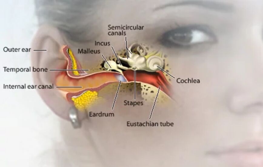
Ears
Ears consist of outer ear, middle ear and inner ear.
Outer ear…
Outer ear is pinna also called auricle. Auditory cannal. There is folds and grieves in it. The fibrous cartilaginous mussle is the muscle with which it is made of. This is elastic fiber bones. This collects all sounds and send to middle ear through auditory cannal.
Middle Ear…
Diaphragm like membrane , also called ear drum separates outer ear from inner ear.
MIS: malleus incus steppus are hammer, anvil and stappas shapped oscicles.
Malleus touches ear drum. Incus articulates with malleus and stapus.
When eardrum vibrates , the malleus vibrates and incus articulates it with stappus.
Fluid in inner ear passes sounds to sound recepters in inner ear .
Inner ear...
Inner ear contain 3 semi circular cannals lying perpendicular to each other.
This helps balancing function of ear. This cannal is connected to cochlea which is spiral muscle sending earing information through rescepters by means of vestigial tube to brain, sound is understood.
Eustachian tube…
Eustachian tube connects inner ear to nasal cavity. Nasal air and auditory air together maintain pressure on the ear making hearing possible.
Sebasius gland…
Sebasius gland makes wax protects ear from insects.
Caicum…
Caicum is the hollow path into auditory canal.
Cartialegenus fluid…
Cartilaginous fluid is filled in inner ear makes earing possible.
Nose…
Nose, the prominent structure between the eyes that serves as the entrance to the respiratory tract and contains the olfactory organ. It provides air for respiration, serves the sense of smell, conditions the air by filtering, warming, and moistening it, and cleans itself of foreign debris extracted from inhalations.
Parts of Nose
- External meatus. Triangular-shaped projection in the center of the face.
- External nostrils. Two chambers divided by the septum.
- Septum. Made up mainly of cartilage and bone and covered by mucous membranes.
- Nasal passages.
- Sinuses.
What are the 5 functions of the nose?
Your nose is involved in several important bodily functions:
- Allows air to enter your body.
- Contributes to how you look and how you sound when you speak.
- Filters and cleans air to remove particles and allergens.
- Provides a sense of smell.
- Warms and moistens air so it can move comfortably into your respiratory system.
What is tip of nose called?
The apex is the tip of the nose. On either side of the apex, the nostrils are formed by the alae (singular = ala). An ala is a cartilaginous structure that forms the lateral side of each naris (plural = nares), or nostril opening. The philtrum is the concave surface that connects the apex of the nose to the upper lip.
What are the little bones in your nose called?
The nasal bones are two small oblong bones, varying in size and form in different individuals; they are placed side by side at the middle and upper part of the face and by their junction, form the bridge of the upper one third of the nose. Each has two surfaces and four borders.
How does a nose work?
Air comes into the body through the nose. As it passes over the specialized cells of the olfactory system, the brain recognizes and identifies smells. Hairs in the nose clean the air of foreign particles. As air moves through the nasal passages, it is warmed and humidified before it goes into the lungs.
Are there veins in your nose?
Facial veins, or broken capillaries, usually appear on the nose, chin, and cheeks. While facial veins typically don’t cause pain or discomfort, they may make you self-conscious. State-of-the-art facial vein treatments can eliminate facial veins and rejuvenate your skin.
What is Cartilage of Septum?
Nasal bridge is the bony part of the nose, overlying the nasal bones, above the part in blue labeled “Cartilage of Septum”. The bridge is between the eyes, and just below them. The lower half of the nose is below the bridge.
What are three functions of the nose?
It provides air for respiration, serves the sense of smell, conditions the air by filtering, warming, and moistening it, and cleans itself of foreign debris extracted from inhalations.
How do nosebleeds happen?
A nosebleed happens when one of the blood vessels in the lining of the nose bursts. Nosebleeds may be caused by infection, injury, allergic reaction, nose picking or an object being pushed into the nostril. Another name for nosebleed is epistaxis. Bleeding from the nose is common in children and is usually not serious.
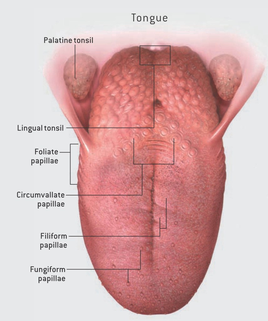
Tongue
The tongue is a muscular organ in the mouth of a typical vertebrate. It manipulates food for mastication and swallowing as part of the digestive process, and is the primary organ of taste. The tongue’s upper surface (dorsum) is covered by taste buds housed in numerous lingual papillae.
Healthy tongues are light pink with some white on the surface.
What are the 5 parts of the tongue?
- Epithelium. The epithelium comprises papillae and taste buds. …
- Muscles. The tongue muscles are voluntary and contain cross-striated muscular fibres.
- Glands. The tongue consists of small and scattered glands. …
- Nerve Supply. …
- Mastication. …
- Deglutition. …
- Taste. …
- Speech.
- What are the 4 types of papillae? The dorsal surface of the mammalian tongue is covered with four kinds of papillae, fungiform, circumvallate, foliate and filiform papillae. With the exception of the filiform papillae, these types of papillae contain taste buds and are known as the gustatory papillae.
- Fungiform papillae are mushroom-shaped structures located on the dorsum of the anterior two-thirds of the tongue.1–3 Due to their rich capillary network, larger size and patchy distribution, fungiform papillae can be identified as reddish dots that contrast to the smaller and more numerous filiform papillae.
- Circumvallate papillae: Also known as vallate papillae, 7-11 of these are located on the backside of your tongue, containing over 100 taste buds each. Fungiform papillae: Over 200 are found on the front side of your tongue and contain 3-5 taste buds each.
- The foliate papillae are leaf shaped with taste buds on the side of the papillae, and these are along the border. The circumvallate papillae contain taste buds along the sides of whorls and are located in the posterior third of the tongue in the shape of a V.
- Filiform papillae are the most numerous of the lingual papillae. They are fine, small, cone-shaped papillae covering most of the dorsum of the tongue. They are responsible for giving the tongue its texture and are responsible for the sensation of touch. … They cover most of the front two-thirds of the tongue’s surface.
2.What are Muscles of tongue?
- The superior longitudinal lingual muscle, which shortens the tongue and curls it upward.
- The inferior longitudinal lingual muscle, which shortens the tongue and curls it downward.
- The transverse lingual muscle, which elongates and narrows the tongue.
- The vertical lingual muscle, which flattens the tongue.
3. What are the 3 Glands?The three main pairs of salivary glands are the parotid glands, the sublingual glands, and the submandibular glands.
- What is the function of parotid gland?The primary function of the parotid gland is the creation of saliva. It’s the saliva itself that performs a number of crucial functions. Saliva is a hypotonic solution created through a joint effort by all the salivary glands.
- The sublingual glands are the smallest of the major salivary glands. These almond-shaped structures are located under the floor of the mouth and below either side of the tongue.
- The submandibular gland is the second largest of the three main salivary glands, which also include the parotid and sublingual glands. The submandibular glands are paired major salivary glands that lie in the submandibular triangle. The glands have a superficial and deep lobe separated by the mylohyoid muscle
4.What are nerve supply to tongue?
Sensory supply:- Anterior two-thirds: Lingual nerve (a branch of the mandibular division of the trigeminal nerve – V3) Posterior one-third: Glossopharyngeal nerve (cranial nerve IX), plus a small branch of the internal laryngeal nerve (branch of the vagus nerve, cranial nerve X).
5. What do you mean by mastication?
1 : to grind or crush (food) with or as if with the teeth : chew The cows were masticating their food. 2 : to soften or reduce to pulp by crushing or kneading. intransitive verb.3 : chew. Other Words from masticate Synonyms More Example Sentences Learn More About masticate.
6. Deglutition?
What are the 3 stages of deglutition?Deglutition is divided into three phases: oropharyngeal, esophageal, and gastroesophageal. The oropharyngeal phase is controlled by the trigeminal (CN V), facial (CN VII), glossopharyngeal (CN IX), vagus (CN X), and hypoglossal nerves (CN XII).
What is deglutition and its process?Swallowing, or deglutition, is a complex reflex mechanism by which food is pushed from the oral cavity into the esophagus and then pushed to the stomach. This movement of food from the oral cavity on to the esophagus and stomach by pushing is called propulsion, and it is an important part of the digestive process.
7. Taste?
What are the tastes of the tongue?There are five universally accepted basic tastes that stimulate and are perceived by our taste buds: sweet, salty, sour, bitter and umami.
8.Speeech?
What are the 4 types of speech?The four basic types of speeches are: to inform, to instruct, to entertain, and to persuade. These are not mutually exclusive of one another. You may have several purposes in mind when giving your presentation. For example, you may try to inform in an entertaining style.
What is an unhealthy tongue? When a tongue is unhealthy. One of the first noticeable symptoms of an unhealthy tongue is a significant change in color from the normal pink shade you’re used to seeing. Other signs of concern can include pain when eating, drinking, and swallowing, as well as new lumps and bumps.
Teeth
Humans usually have 20 primary (deciduous, “baby” or “milk”) teeth and 32 permanent (adult) teeth. Teeth are classified as incisors, canines, premolars (also called bicuspids), and molars. Incisors are primarily used for cutting, canines are for tearing, and molars serve for grinding.
We have four different types of teeth, with each type serving a particular purpose for eating and chewing.
- 8 Incisors.
- 4 Canines.
- 8 Premolars.
- 12 Molars (including 4 wisdom teeth)
What are incisors? Your eight incisor teeth are located in the front part of your mouth. You have four of them in your upper jaw and four in your lower jaw. Incisors are shaped like small chisels.
The last sets of baby teeth to go are the canines and primary second molars. The canines are usually lost between the ages of 9 and 12 years old, while the primary second molars are the last baby teeth that your child will lose. These final sets of teeth are usually shed between the ages of 10 and 12.
Symptoms of molar comming in?
- Your child may be drooling more than usual.
- They might be unusually irritable.
- Your child may be chewing on their fingers, clothing, or toys.
- They might have a consistent low-grade temperature of about 99 degrees F.
- If you’re able to get a look – they have red gums at the eruption zone.
- Interrupted sleep.
Are your molars permanent?These important teeth sometimes are mistaken for pri- mary teeth. However, they are permanent and must be cared for properly if they are to last throughout the child’s lifetime. The six-year molars also help determine the shape of the lower face and affect the position and health of other permanent teeth.
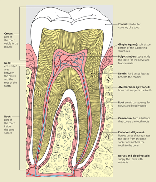
Tooth
A tooth (plural teeth) is a hard, calcified structure found in the jaws (or mouths) of many vertebrates and used to break down food.
Your teeth are composed of four dental tissues. Three of them—enamel, dentin and cementum—are hard tissues. The fourth tissue—pulp, or the center of the tooth that contains nerves, blood vessels and connective tissue—is a soft, or non-calcified, tissue.
- Enamel. Hard calcified tissue covering the dentin in the crown of tooth. Because it contains no living cells, tooth enamel cannot repair damage from decay or from wear. Only a dentist can correct these conditions.
- Anatomical Crown. The visible part of your tooth. It is normally covered by enamel.
- Gums (also called gingiva.) Soft tissues that cover and protect the roots of your teeth and cover teeth that have not yet erupted.
- Pulp Chamber. The space occupied by the pulp—the soft tissue at the center of your teeth containing nerves, blood vessels and connective tissue.
- Neck. The area where the crown joins the root.
- Dentin. That part of the tooth that is beneath enamel and cementum. It contains microscopic tubules (small hollow tubes or canals). When dentin loses its protective covering (enamel), the tubules allow heat and cold or acidic or sticky foods to stimulate the nerves and cells inside the tooth, causing sensitivity.
- Jawbone (Alveolar Bone.) The part of the jaw that surrounds the roots of the teeth.
- Root Canal. The portion of the pulp cavity inside the root of a tooth; the chamber within the root of the tooth that contains the pulp.
- Cementum. Hard connective tissue covering the tooth root, giving attachment to the periodontal ligament.
- Periodontal Ligament. A system of collagenous connective tissue fibers that connect the root of a tooth to its socket.
Bones
What are Bones?Bones provide the structure for our bodies. The adult human skeleton is made up of 206 bones. These include the bones of the skull, spine (vertebrae), ribs, arms and legs. Bones are made of connective tissue reinforced with calcium and specialised bone cells.
What is bone and its function?Bones have many functions. They support the body structurally, protect our vital organs, and allow us to move. Also, they provide an environment for bone marrow, where the blood cells are created, and they act as a storage area for minerals, particularly calcium.
There are four different types of bone in the human body:
- Long bone – has a long, thin shape. …
- Short bone – has a squat, cubed shape. …
- Flat bone – has a flattened, broad surface. …
- Irregular bone – has a shape that does not conform to the above three types.
Types of Bones and their function:-
The Long and the Short of It: The Five Types of Bones
- Flat Bones Protect Internal Organs. …
- Long Bones Support Weight and Facilitate Movement. …
- Short Bones Are Cube-shaped. …
- Irregular Bones Have Complex Shapes. …
- Sesamoid Bones Reinforce Tendons.
Skin
The skin is the largest organ of the body, with a total area of about 20 square feet. The skin protects us from microbes and the elements, …
The skin is the body’s largest organ. It covers the entire body. It serves as a protective shield against heat, light, injury, and infection. The skin also: Regulates body temperature.
What is skin and function?
Provides a protective barrier against mechanical, thermal and physical injury and hazardous substances. Prevents loss of moisture. Reduces harmful effects of UV radiation. Acts as a sensory organ (touch, detects temperature).
What are the seven most important layers of your skin?
- Stratum corneum.
- Stratum lucidum.
- Stratum granulosum.
- Stratum spinosum.
- Stratum basale.
- Dermis.
- Hypodermis.
What is skin made of?
Skin has three layers: The epidermis, the outermost layer of skin, provides a waterproof barrier and creates our skin tone. The dermis, beneath the epidermis, contains tough connective tissue, hair follicles, and sweat glands. The deeper subcutaneous tissue (hypodermis) is made of fat and connective tissue.
What is the 5 layers of the epidermis?The layers of the epidermis include the stratum basale (the deepest portion of the epidermis), stratum spinosum, stratum granulosum, stratum lucidum, and stratum corneum (the most superficial portion of the epidermis).
What are the 5 types of cells found in the epidermis?Terms in this set (5)
- Stem Cells. Undifferentiated cells give rise to keratinocytes, deepest layer of epidermis (stratum basale)
- Keratinocytes. Great majority of epidermal cells, synthesize keratine.
- Melanocytes. …
- Tactile (Merkel) Cells. …
- Dendritic (Langerhans) Cells.
What are the 7 main functions of the skin?
Storing lipids (fats) and water. Creating sensation through nerve endings that detect temperature, pressure, vibration, touch, and injury. Controlling water loss by preventing water from escaping by evaporation. Providing water resistance by preventing nutrients from being washed from the skin.
Where is the thickest skin?
palms of the handsEpidermis varies in thickness throughout the body depending mainly on frictional forces and is thickest on the palms of the hands and soles of the feet, and thinnest in the face (eyelids) and genitalia.
What is Skin cycle?
A skin cycle is the process where a new skin cell is formed at the deepest layer of the epidermis and works it’s way up to the surface of the skin. … On average a skin cycle is 5-6 weeks. At the age of 19-21,the process can take 14-21 days compared to a middle-aged adult where it is estimated to be 28 days.
What vitamin does your skin produce?
The skin is responsible for producing vitamin D. During exposure to sunlight, ultraviolet radiation penetrates into the epidermis and photolyzes provitamin D3 to previtamin D3.
What does Skin secrete?
Skin secretions originate from glands that in dermal layer of the epidermis. Sweat, a physiological aid to body temperature regulation, is secreted by eccrine glands. Sebaceous glands secrete the skin lubricant sebum. Sebum is secreted onto the hair shaft and it prevents the hair from splitting.
Which vitamin tablet is good for skin?
Vitamin D is one of the best vitamins for your skin, along with vitamins C, E, and K. Making sure you get enough vitamins can keep your skin looking healthy and youthful.
What cells are in skin?
The epidermis has three main types of cell: Keratinocytes (skin cells) Melanocytes (pigment-producing cells) Langerhans cells (immune cells).
Is vitamin E or C better for skin?
Vitamin C is an antioxidant and slows the rate of free-radical damage to collagen that can contribute to dry skin, fine lines and wrinkles. Vitamin E is an antioxidant that protects and repairs your skin and can help prevent premature aging of your skin and damage to your DNA.
(DER-mis) The inner layer of the two main layers of the skin. The dermis has connective tissue, blood vessels, oil and sweat glands, nerves, hair follicles, and other structures. It is made up of a thin upper layer called the papillary dermis, and a thick lower layer called the reticular dermis.
What is dermis and its function?The dermis is a fibrous structure composed of collagen, elastic tissue, and other extracellular components that includes vasculature, nerve endings, hair follicles, and glands. The role of the dermis is to support and protect the skin and deeper layers, assist in thermoregulation, and aid in sensation
Hair
What is an hair?
Hair is a protein filament that grows from follicles found in the dermis. Hair is one of the defining characteristics of mammals. … Most common interest in hair is focused on hair growth, hair types, and hair care, but hair is also an important biomaterial primarily composed of protein, notably alpha-keratin.
What are the 5 types of hair?
- Straight hair.
- Wavy hair.
- Curly hair.
- Coily hair.
What are the hair types?
The four hair types are type 1 straight, type 2 wavy, type 3 curly and type 4 tight curls. Hair type and texture are determined by several factors including genetics. Straight hair is one of the most common hair types across the world. Each person has a unique texture.
https://www.healthline.com/health/beauty-skin-care/types-of-hair
Finger nails
A nail is a claw-like plate at the tip of the fingers and toes in most primates. Nails correspond to claws found in other animals. Fingernails and toenails are made of a tough protective protein called alpha-keratin, which is a polymer.
How do you treat finger nails?Fingernail care: Do’s
- Keep fingernails dry and clean. This prevents bacteria from growing under your fingernails. …
- Practice good nail hygiene. Use a sharp manicure scissors or clippers. …
- Use moisturizer. …
- Apply a protective layer. …
- Ask your doctor about biotin.
Nutrients to keep your nails healthy.
- Biotin.
- Other B Vitamins. Other B vitamins are also important for nail health. …
- Iron. …
- Magnesium. …
- Protein. …
- Omega-3 Fatty Acids. …
- Vitamin C. …
- Zinc.
https://lesalon.com/blog/dip-nails-and-acrylics/
Human Face
Why is the human face so important?
The face is the “organ of emotion,” and we constantly read facial expressions to understand what others are feeling. … The face is one of our most important possessions. It has been called “the organ of emotion” and, indeed, the face provides vital clues to our own feelings and those of the people around us.
The 7 basic face shapes are oval, round, square, diamond, heart, pear and oblong.
The front of the human head is called the face. It includes several distinct areas, of which the main features are: The forehead, comprising the skin beneath the hairline, bordered laterally by the temples and inferiorly by eyebrows and ears. The eyes, sitting in the orbit and protected by eyelids and eyelashes.
The face can be divided into five main parts: the forehead, eyes, cheeks, nose, and perioral area.
What is a perioral area?
The ‘perioral’ area of your face is the area immediately surrounding your mouth. The perioral area includes several features: both lips, upper lip area under the nose, nasiolabial folds (“smile lines” or “laugh lines”), and the corners of the mouth.
According to its very specific definitions, the perfect face included these key features: Length of the face equals the length of three noses. Width of an eye in between the eyes. Upper and lower lips are the same width. Symmetrical eyebrows conforming to the line of the nose.
The amazing variety of human faces – far greater than that of most other animals – is the result of evolutionary pressure to make each of us unique and easily recognizable, according to a new study by University of California, Berkeley, scientists.
Age related facial remedies…
What is perioral rejuvenation?
Essentially, perioral rejuvenation is the restoration of your lips and mouth area. In particular, some of the age-related concerns that can be corrected with this treatment include: Lipstick lines. Smoker’s lines. Marionette lines.
Lips…
1. The color of your lips is caused by visible blood capillaries under your skin. They are visible because the lips have one of the thinnest layers of skin on the body.
2. They are the most sensitive part on your body-they have over 1 million nerve endings.
3.Lips never sweat- because they do not have sweat glands.
4. They get thinner as you get older.
5.They are unique- like finger prints.
6.They are the only parts of the body where the inside extends to the outside. The membrane that makes up the inner lips also makes the outer lips.
7.The dip above your upper lip is called the Philtrum
(home)
Hands
A hand is a prehensile, multi-fingered appendage located at the end of the forearm or forelimb of primates such as humans, chimpanzees, monkeys, and lemurs.
A hand is the part of the body at the end of an arm. … The five bones inside this part of the hand are called metacarpals. The wrist connects the hand to the arm. The hand has 27 bones including the wrist bones. When the fingers are all bent tightly, the hand forms a fist.
What is so special about hands?–The hands have 29 major joints, at least 123 ligaments, 34 muscles, 48 nerves and 30 arteries. … –Human hands are able to make grips that other primates, such as chimps and gorillas, cannot. This is because we have shorter hands and longer, more powerful thumbs then our primate relatives.
Parts of a Hand
- Bones are hard tissues that give your hand shape and stability.
- Phalanges are the finger bones.
- Metacarpals are the middle part of the hand bones.
- Carpals are the wrist bones.
- Joints are places where bones fit together, allowing movement.
What are fingers called?Phalanges. The 14 bones that are found in the fingers of each hand and also in the toes of each foot. Each finger has 3 phalanges (the distal, middle, and proximal); the thumb only has 2.
What is the palm of your hand?The palm comprises the underside of the human hand. Also known as the broad palm or metacarpus, it consists of the area between the five phalanges (finger bones) and the carpus (wrist joint).
Legs
The human leg, in the general word sense, is the entire lower limb of the human body, including the foot, thigh and even the hip or gluteal region. However, the definition in human anatomy refers only to the section of the lower limb extending from the knee to the ankle, also known as the crus or, especially in non-technical use, the shank. Legs are used for standing, and all forms of locomotion including recreational such as dancing, and constitute a significant portion of a person’s mass. Female legs generally have greater hip anteversion and tibiofemoral angles, but shorter femur and tibial lengths than those in males.
In human anatomy, the lower leg is the part of the lower limb that lies between the knee and the ankle. The thigh is between the hip and knee and makes up the rest of the lower limb. The term lower limb or “lower extremity” is commonly used to describe all of the leg. This article generally follows the common usage.
The leg from the knee to the ankle is called the crus or cnemis /ˈniːmɪs/. The calf is the back portion, and the tibia or shinbone together with the smaller fibula make up the front of the lower leg.
